. 2010 Jul;5(7):545-51.
doi: 10.1038/nnano.2010.104. Epub 2010 Jun 13.

Subcellular-resolution delivery of a cytokine through precisely manipulated nanowires

Affiliations

Subcellular-resolution delivery of a cytokine through precisely manipulated nanowires

Donglei Fan et al. Nat Nanotechnol. 2010 Jul.

Abstract

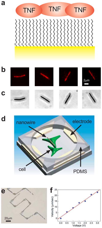
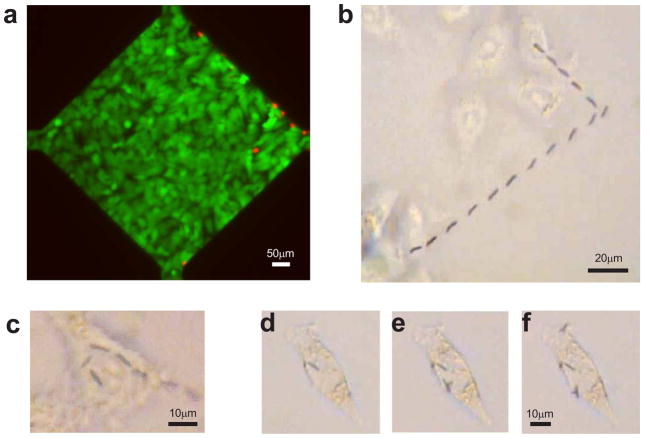
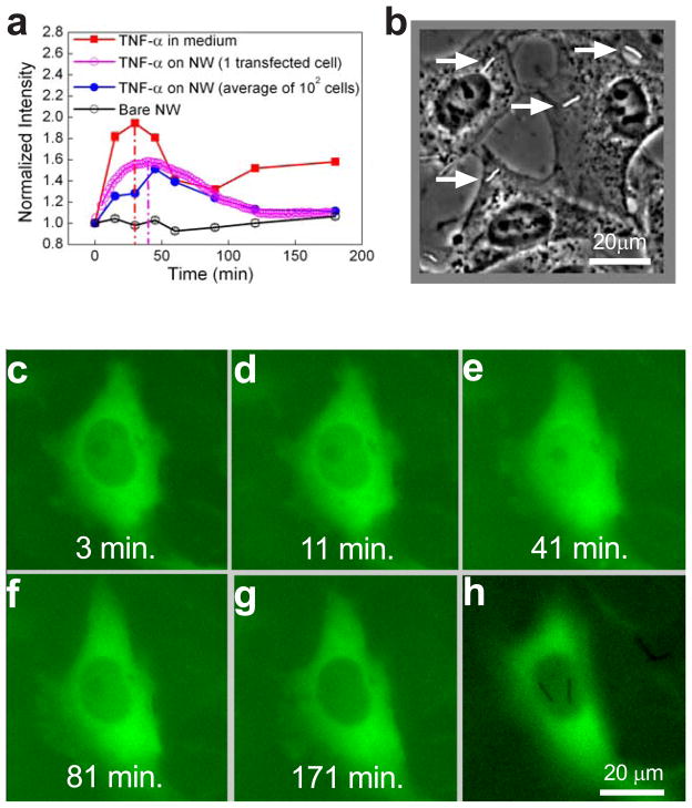
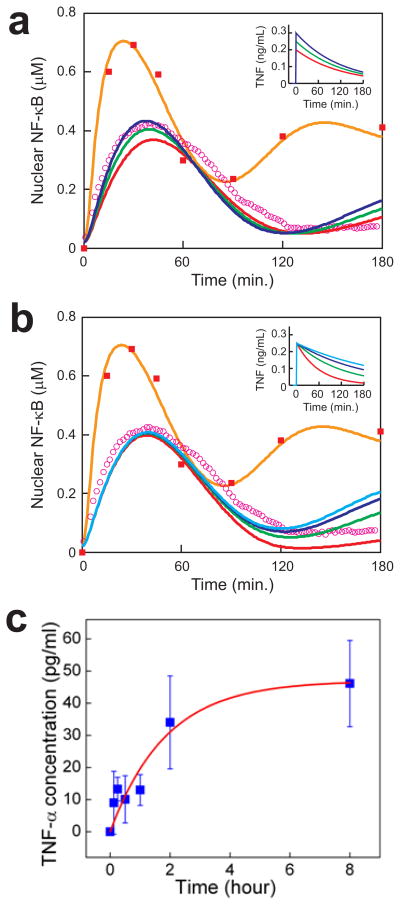
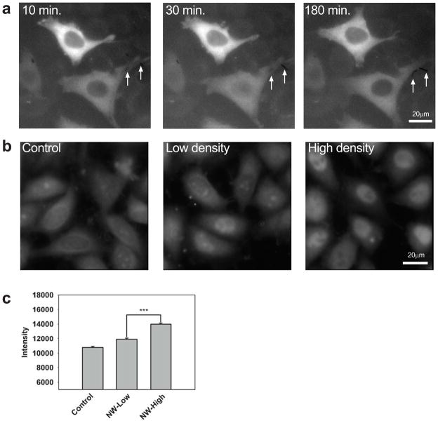
Precise delivery of molecular doses of biologically active chemicals to a pre-specified single cell among many, or a specific subcellular location, is still a largely unmet challenge hampering our understanding of cell biology. Overcoming this could allow unprecedented levels of cell manipulation and targeted intervention. Here, we show that gold nanowires conjugated with a cytokine such as tumour-necrosis factor-alpha can be transported along any prescribed trajectory or orientation using electrophoretic and dielectrophoretic forces to a specific location with subcellular resolution. The nanowire, 6 microm long and 300 nm in diameter, delivered the cytokine and activated canonical nuclear factor-kappaB signalling in a single cell. Combined computational modelling and experimentation indicated that cell stimulation was highly localized to the nanowire vicinity. This targeted delivery method has profound implications for controlling signalling events on the single cell level.


Figures

Figure 1
Figure 1
Nanowire functionalization and movement. a, Schematic shows nanowire (yellow) surface modified with 1-dodecanethiol, rendering the surface hydrophobic and allowing the adsorption of TNF α (red). Fluorescent (b) and phase contrast (c) images of rhodamine-conjugated TNF α adsorbed on the surface of individual nanowires. d, Schematic shows two pairs of parallel electrodes that control nanowire orientation and movement by alternating and constant electric fields. PDMS placed over the electrodes forms a well in which cells are cultured. e, Overlay of a time series of phase contrast images showing the trajectory of the nanowires through precise rotations and translations (see also Supplementary Movie 1). f, Nanowire transport speed is proportional to the applied constant electric field, allowing precise control over nanowire velocity.
Figure 2
Figure 2
Nanowire delivery to pre-selected cells. a, Live (green) and dead (red) cell staining showing that typical voltages used to manipulate nanowires does not affect the viability of most cells in the operating area. Dead cells were confined to the areas immediately adjacent to the electrodes. b, Overlay of a time series of phase contrast images showing delivery of a single nanowire to the cell at the top of the image. (See also Supplementary Movie 2.) c, Overlay of a time series of phase contrast images showing delivery of a single nanowire to sub-divisions of the same cell. (See also Supplementary Movie 3.) d–f, Phase contrast images showing sequential delivery of nanowires to a single cell.
Figure 3
Figure 3
Delivery of functional TNF α by nanowires. a, Average time-course of nuclear NF-κB concentration in wildtype HeLa cells exposed to 10ng/mL TNF α dissolved in the medium (red), TNF α-coated nanowires (blue), or bare nanowires (black) as measured by immunocytochemistry. Nuclear NF-κB measured in a HeLa cell expressing p65-GFP exposed to TNF α-coated nanowires is shown in magenta. b, Phase contrast image showing TNF α-coated nanowires (arrows) at a concentration that yielded ~1 nanowire per cell. c–h, Representative HeLa cell expressing p65-GFP stimulated with TNF α-coated nanowires for the indicated durations (panels c–g). Nuclear p65 levels peak around 40 min (see also Supplementary Movie 4). In panel h, the same cell is imaged at a different focal plane to show the two TNF α-coated nanowires that contact and stimulate the cell.
Figure 4
Figure 4
Simulations predict localized TNF α delivery. a,b, The simulated response to 10ng/mL TNF α (orange) compares well with experiment (red squares, same as Fig. 3a). Simulated responses to TNF α inputs of the form c(t) = cos exp(−t/tos) (t > 0) are shown. In panel a, tos = 120 min. and cos = 0.20 (red), 0.25 (green), 0.30 (blue) ng/mL. In panel b, cos = 0.25 ng/mL and tos = 60 (red), 120 (green), 180 (blue), 240 (light blue) min. The green curves give the best fit to experiment (magenta circles, same as Fig. 3a). See Methods for the model description. c, TNF α-desorption kinetics determined by ELISA measurements of TNF α concentration in the supernatant of a solution of 5×10⁶ nanowires/mL. See text for description of the fitted exponential curve (red).
Figure 5
Figure 5
Selective stimulation by TNF α-coated nanowires. a, HeLa cells expressing p65-GFP at the indicated times following stimulation with TNF α-coated nanowires. The lower cell contacts two nanowires (arrows) and exhibits p65 nuclear translocation whereas the upper cell contacts no nanowires and exhibits no translocation. (See also Supplementary Movie 5.) b,c, Wildtype HeLa cells stained for NF-κB following exposure to solutions of 0 (control), 0.2 × 10⁶ (low density), or 1 × 10⁶ (high density) TNF α-coated nanowires/mL, corresponding to 0, ~0.3, ~1.5 nanowires/cell on average, respectively. In (b), representative images are shown. In (c), the average NF-κB level is plotted (more than 150 cells measured for each condition). NF-κB levels were significantly different (p < .001) between the high and low stimulation cases.

Comment in

References

    1. Bray SJ. Notch signalling: a simple pathway becomes complex. Nat Rev Mol Cell Biol. 2006;7:678–89. - PubMed
    1. Delves PJ, Roitt IM. The immune system. First of two parts. N Engl J Med. 2000;343:37–49. - PubMed
    1. Nagata S. Fas ligand-induced apoptosis. Annu Rev Genet. 1999;33:29–55. - PubMed
    1. Wu YI, Frey D, Lungu OI, Jaehrig A, Schlichting I, et al. A genetically encoded photoactivatable Rac controls the motility of living cells. Nature. 2009;461:104–8. - PMC - PubMed
    1. Lee HM, Larson DR, Lawrence DS. Illuminating the chemistry of life: design, synthesis, and applications of “caged” and related photoresponsive compounds. ACS Chem Biol. 2009;4:409–27. - PMC - PubMed

Publication types

MeSH terms