

Deep anterior lamellar keratoplasty: indications, surgical techniques and complications
- PMID: 20543934
- PMCID: PMC2880371
- DOI: 10.4103/0974-9233.61214
Deep anterior lamellar keratoplasty: indications, surgical techniques and complications
Abstract
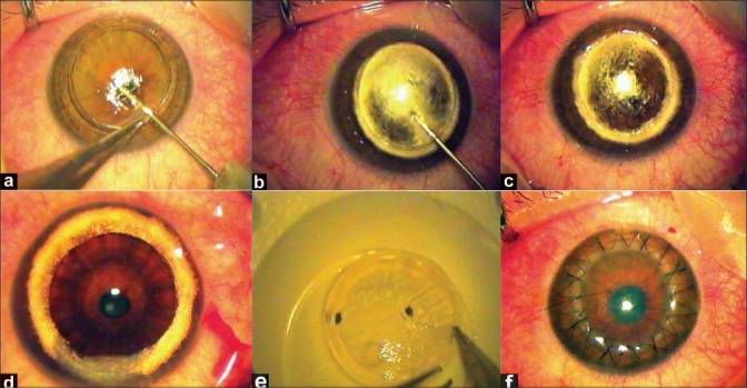
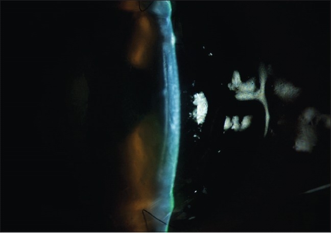
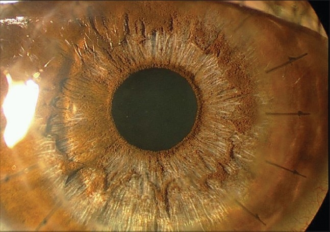
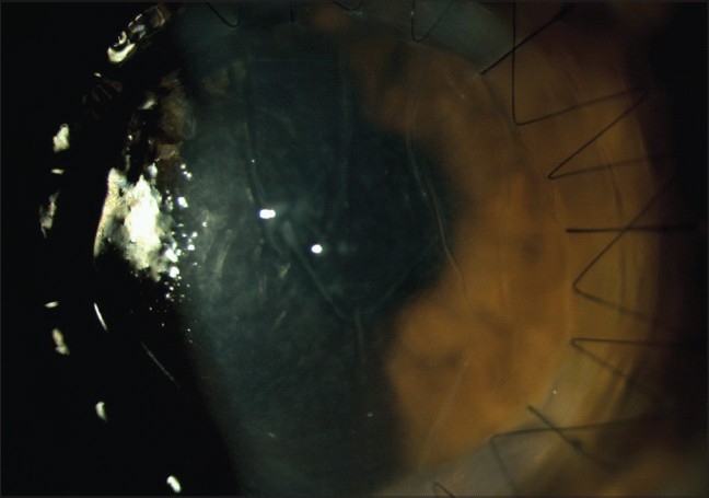
The concept of lamellar keratoplasty (LK) is not new. However, it had been abandoned and largely replaced by the time-honored technique of penetrating keratoplasty (PK) because LK is technically demanding, time consuming and gives suboptimal visual outcomes due to interface irregularity arising from manual lamellar dissection. Recent improvements in surgical instruments and introduction of new techniques of maximum depth of corneal dissection as well as inherent advantages such as preservation of globe integrity and elimination of endothelial graft rejection have resulted in a re-introduction of LK as an acceptable alternative to conventional PK. This review article describes the indications, different techniques, clinical outcomes and complications of deep anterior LK.
Keywords: Anwar's Big-bubble Technique; Deep Anterior Lamellar Keratoplasty; Lamellar Keratoplasty; Melles'Technique; Penetrating Keratoplasty.
Conflict of interest statement
Figures
Similar articles
-
Lamellar keratoplasty: a literature review.J Ophthalmol. 2013;2013:894319. doi: 10.1155/2013/894319. Epub 2013 Oct 7. J Ophthalmol. 2013. PMID: 24223301 Free PMC article. Review.
-
Future directions in lamellar corneal transplantation.Cornea. 2007 Oct;26(9 Suppl 1):S21-8. doi: 10.1097/ICO.0b013e31812f685c. Cornea. 2007. PMID: 17881911 Review.
-
Lamellar keratoplasty techniques.Indian J Ophthalmol. 2018 Sep;66(9):1239-1250. doi: 10.4103/ijo.IJO_95_18. Indian J Ophthalmol. 2018. PMID: 30127133 Free PMC article. Review.
-
Bubbling With Confidence: A Study of Numerous Deep Anterior Lamellar Keratoplasty Cases Using Anwar's Big Bubble Technique.Cureus. 2023 Oct 5;15(10):e46528. doi: 10.7759/cureus.46528. eCollection 2023 Oct. Cureus. 2023. PMID: 37927665 Free PMC article.
-
Comparative study of keratoconus between Anwar's deep anterior lamellar keratoplasty versus converted penetrating keratoplasty.Arch Soc Esp Oftalmol. 2015 Jun;90(6):257-63. doi: 10.1016/j.oftal.2014.11.008. Epub 2015 Mar 25. Arch Soc Esp Oftalmol. 2015. PMID: 25817955 English, Spanish.
Cited by
-
Therapeutic effect of deep anterior lamellar keratoplasty for active or quiescent herpetic stromal keratitis.Graefes Arch Clin Exp Ophthalmol. 2012 Aug;250(8):1187-94. doi: 10.1007/s00417-012-1947-2. Epub 2012 Feb 17. Graefes Arch Clin Exp Ophthalmol. 2012. PMID: 22349979
-
Management of Corneal Clouding in Patients with Mucopolysaccharidosis.J Clin Med. 2021 Jul 24;10(15):3263. doi: 10.3390/jcm10153263. J Clin Med. 2021. PMID: 34362047 Free PMC article. Review.
-
Using Artificial Intelligence for an Efficient Prediction of Outcomes of Deep Anterior Lamellar Keratoplasty (DALK) in Advanced Keratoconus.Transl Vis Sci Technol. 2025 May 1;14(5):30. doi: 10.1167/tvst.14.5.30. Transl Vis Sci Technol. 2025. PMID: 40434371 Free PMC article.
-
Pediatric keratoconus - Current perspectives and clinical challenges.Indian J Ophthalmol. 2021 Feb;69(2):214-225. doi: 10.4103/ijo.IJO_1263_20. Indian J Ophthalmol. 2021. PMID: 33463562 Free PMC article. Review.
-
Low-temperature vacuum evaporation as a novel dehydration process for the long-term preservation of transplantable human corneal tissue.Cell Tissue Bank. 2025 Jan 9;26(1):7. doi: 10.1007/s10561-024-10155-y. Cell Tissue Bank. 2025. PMID: 39779599 Free PMC article.
References
-
- Watson SL, Ramsay A, Dart JK, Bunce C, Craig E. Comparison of deep lamellar keratoplasty and penetrating keratoplasty in patients with keratoconus. Ophthalmology. 2004;111:1676–82. - PubMed
-
- Watson SL, Tuft SJ, Dart JK. Patterns of rejection after deep lamellar keratoplasty. Ophthalmology. 2006;113:556–60. - PubMed
-
- Terry MA. The evolution of lamellar grafting techniques over twenty-five years. Cornea. 2000;19:611–6. - PubMed
-
- Morris E, Kirwan JF, Sujatha S, Rostron CK. Corneal endothelial specular microscopy following deep lamellar keratoplasty with lyophilised tissue. Eye. 1998;12:619–22. - PubMed
-
- Panda A, Bageshwar LM, Ray M, Singh JP, Kumar A. Deep lamellar keratoplasty versus penetrating keratoplasty for corneal lesions. Cornea. 1999;18:172–5. - PubMed
LinkOut - more resources
Full Text Sources
