Review
. 2010 Oct;67(19):3287-97.
doi: 10.1007/s00018-010-0425-4. Epub 2010 Jun 11.

Neuronal ciliary signaling in homeostasis and disease

Affiliations
Review

Neuronal ciliary signaling in homeostasis and disease

Jill A Green et al. Cell Mol Life Sci. 2010 Oct.

Abstract

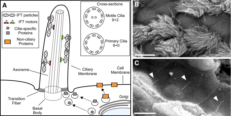
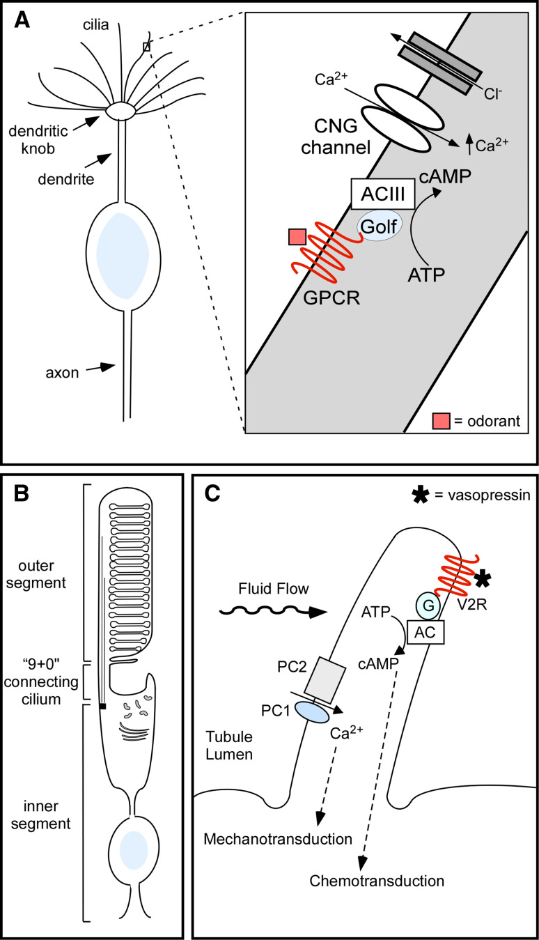
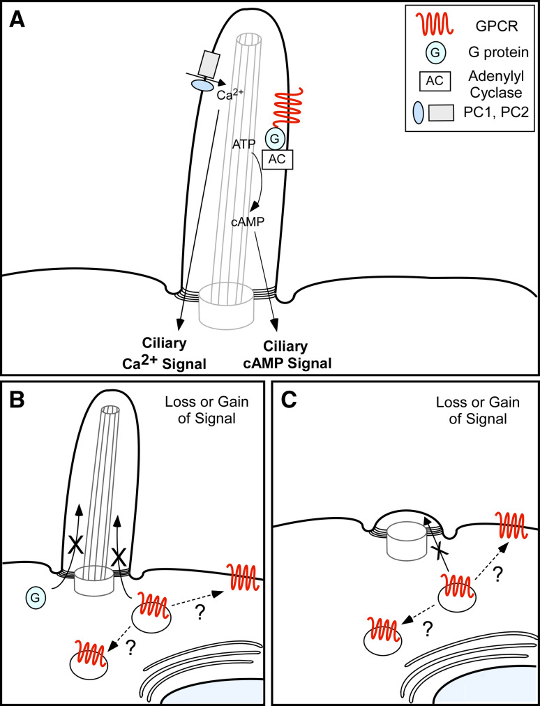
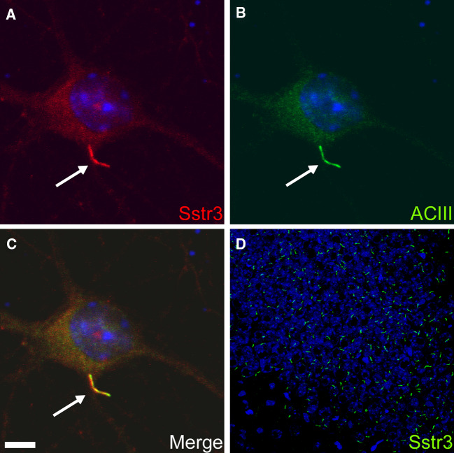
Primary cilia are a class of cilia that are typically solitary, immotile appendages present on nearly every mammalian cell type. Primary cilia are believed to perform specialized sensory and signaling functions that are important for normal development and cellular homeostasis. Indeed, primary cilia dysfunction is now linked to numerous human diseases and genetic disorders. Collectively, primary cilia disorders are termed as ciliopathies and present with a wide range of clinical features, including cystic kidney disease, retinal degeneration, obesity, polydactyly, anosmia, intellectual disability, and brain malformations. Although significant progress has been made in elucidating the functions of primary cilia on some cell types, the precise functions of most primary cilia remain unknown. This is particularly true for primary cilia on neurons throughout the mammalian brain. This review will introduce primary cilia and ciliary signaling pathways with a focus on neuronal cilia and their putative functions and roles in human diseases.


Figures

Fig. 1
Fig. 1
Schematic of cilia structure and examples of motile and primary cilia. a Membrane and cytosolic proteins destined for the ciliary compartment are transported in Golgi-derived vesicles and exocytosed at the base of the cilium where they associate with intraflagellar transport (IFT) particles. The transition fibers form a selective barrier to the ciliary compartment and only proteins containing specific ciliary targeting motifs are allowed access. Following entry into the cilium, proteins are transported along the axoneme. Cross-sections show the typical microtubule structures of motile and primary cilia. b Scanning electron micrograph showing numerous motile cilia protruding from epithelial cells of a mouse trachea. Scale bar 5 μm. c Scanning electron micrograph showing solitary primary cilia (arrowheads) projecting from mouse renal epithelial cells lining the nephron. Scale bar 5 μm
Fig. 2
Fig. 2
Examples of ciliary chemo-, photo-, and mechanotransduction. a Schematic of a single olfactory sensory neuron. Boxed region of interest is magnified and illustrates the ciliary signaling pathway. Odorant activation of olfactory G protein-coupled receptors (GPCRs) results in an increase in cAMP levels, which is mediated by type 3 adenylyl cyclase (ACIII). This results in activation of cyclic nucleotide-gated (CNG) channels leading to an increase in Ca2+ levels, subsequent activation of chloride channels, and depolarization of the neuron. b Schematic of a photoreceptor, which is comprised of an inner and outer segment that are connected by a “9+0” connecting cilium. Proteins are synthesized in the inner segment and transported by IFT across the connecting cilium to the outer segment, which is a highly modified cilium, where they mediate phototransduction. c Schematic of a renal cilium demonstrating Ca2+ signaling mediated by the ciliary proteins polycystin 1 (PC1) and polycystin 2 (PC2) in response to bending of the cilium by fluid flow. Renal cilia may also act as chemosensors by mediating vasopressin activation of the type 2 vasopressin receptor (V2R) on the ciliary membrane, which in turn modulates cAMP-signaling
Fig. 3
Fig. 3
Model of ciliary signaling disruption. a Signaling proteins are enriched in the cilium where they coordinate cilia-specific signaling that is transmitted to the cell. Two examples of ciliary signaling pathways are illustrated; PC1- and PC2-mediated Ca2+ signaling and GPCR-mediated cAMP-signaling. b Disruption in trafficking of signaling proteins (indicated by X) into the ciliary compartment leads to loss of a cilia-specific signal. Ciliary receptors that are not trafficked to the cilium may accumulate in intracellular vesicles or mislocalize on the cell membrane (indicated by ?), possibly resulting in a gain of signal. c Defects in cilia structure prevents proper localization of ciliary signaling proteins (indicated by X) and leads to loss of a cilia-specific signal. Ciliary receptors that are not trafficked to the cilium may accumulate in intracellular vesicles or mislocalize on the cell membrane (indicated by ?), possibly resulting in a gain of signal
Fig. 4
Fig. 4
Localization of somatostatin receptor subtype 3 (Sstr3) to neuronal cilia. a,b Images of a day 7 mouse hippocampal neuron immunolabeled with antibodies to Sstr3 (red) and type 3 adenylyl cyclase (ACIII; green). c Merged image demonstrating colocalization of Sstr3 and ACIII to neuronal cilia. Nuclei were stained with DRAQ5 (blue) and the cilium is indicated with arrows. Scale bar 5 μm. d Adult mouse brain section corresponding to the CA3 region of the hippocampus immunolabeled with an antibody to Sstr3 (green). Note the abundance of Sstr3-positive cilia. Nuclei were stained with DRAQ5 (blue). (All images courtesy of Nicolas Berbari, University of Alabama, Birmingham, AL, USA)

References

    1. Nonaka S, Tanaka Y, Okada Y, Takeda S, Harada A, Kanai Y, Kido M, Hirokawa N. Randomization of left–right asymmetry due to loss of nodal cilia generating leftward flow of extraembryonic fluid in mice lacking KIF3B motor protein. Cell. 1998;95:829–837. doi: 10.1016/S0092-8674(00)81705-5. - DOI - PubMed
    1. Bloodgood RA. Sensory reception is an attribute of both primary cilia and motile cilia. J Cell Sci. 2010;123:505–509. doi: 10.1242/jcs.066308. - DOI - PubMed
    1. Shah AS, Ben-Shahar Y, Moninger TO, Kline JN, Welsh MJ. Motile cilia of human airway epithelia are chemosensory. Science. 2009;325:1131–1134. doi: 10.1126/science.1173869. - DOI - PMC - PubMed
    1. Pedersen LB, Rosenbaum JL. Intraflagellar transport (IFT) role in ciliary assembly, resorption and signalling. Curr Top Dev Biol. 2008;85:23–61. doi: 10.1016/S0070-2153(08)00802-8. - DOI - PubMed
    1. Rosenbaum JL, Witman GB. Intraflagellar transport. Nat Rev Mol Cell Biol. 2002;3:813–825. doi: 10.1038/nrm952. - DOI - PubMed

Publication types

LinkOut - more resources