Review
. 2011 Mar;469(3):776-89.
doi: 10.1007/s11999-010-1414-5.

The radiographic approach to child abuse

Affiliations
Review

The radiographic approach to child abuse

Jerry R Dwek. Clin Orthop Relat Res. 2011 Mar.

Abstract

Background: Osseous injuries are a major facet of child abuse and in most patients radiographic imaging plays a major role in diagnosis. While some injuries are typically produced as a result of excessive and inappropriate force other injuries are nonspecific in terms of their causation, but become suspicious when the history provided by the caretakers is inconsistent with the type of injury produced.

Questions/purposes: I detail the radiographic imaging of the more characteristic of the highly specific injuries, discuss the major issues that relate to some moderate- or low-specificity injuries, and describe several diseases that mimic abuse.

Methods: A review of the current and recent literature focused on the radiographic imaging of child abuse was performed by searching the National Library of Medicine database at pubmed.gov. Keywords used included: radiology, fracture, child abuse, and/or nonaccidental trauma.

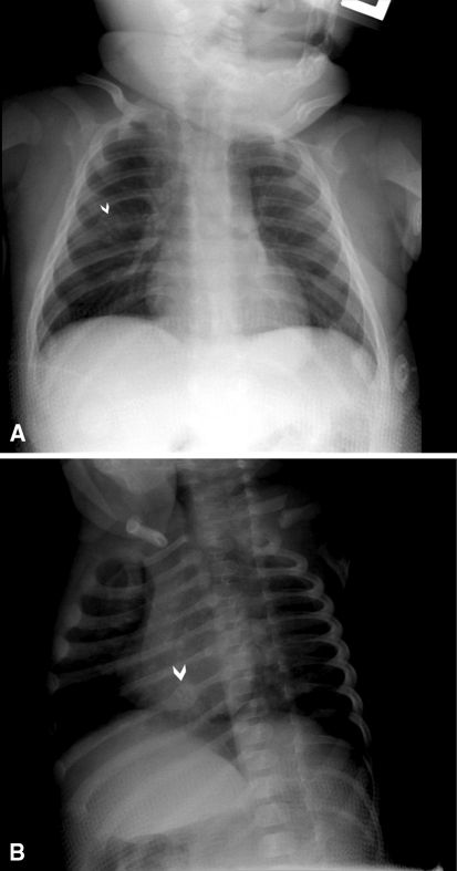
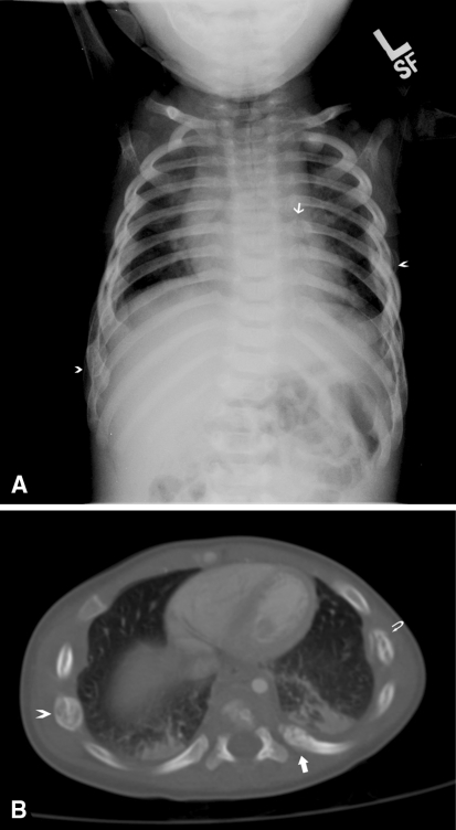
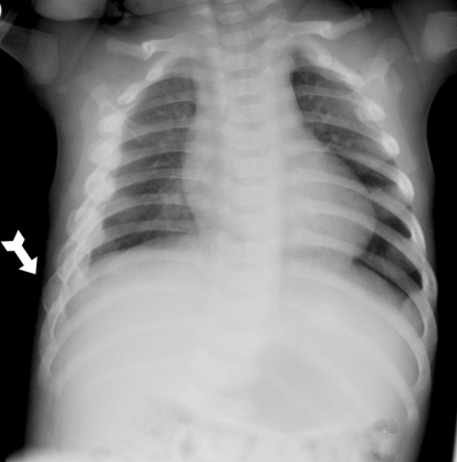
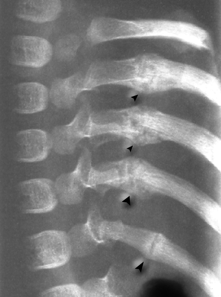
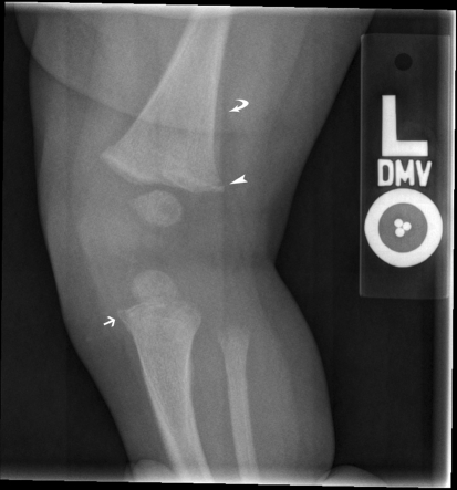
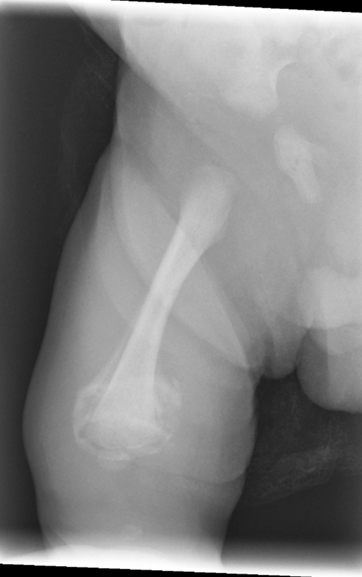
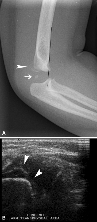
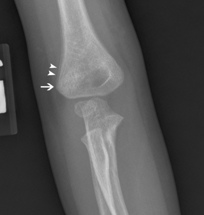
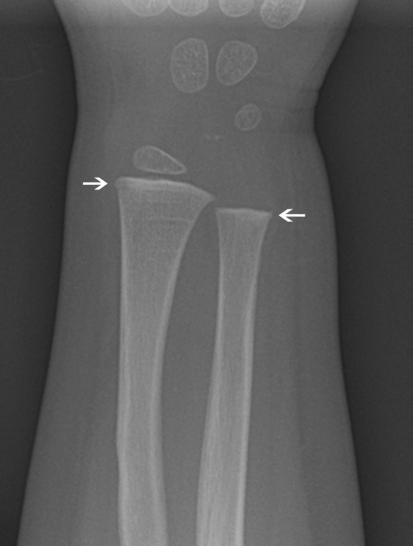
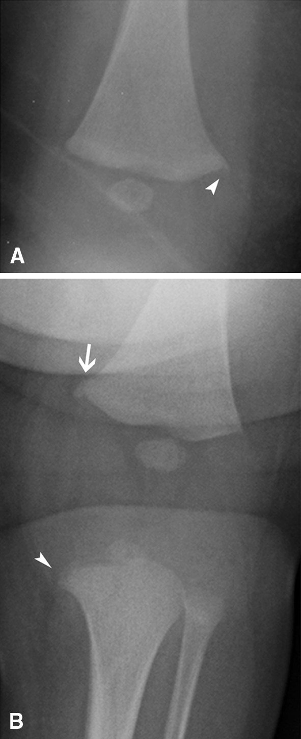
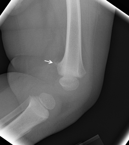
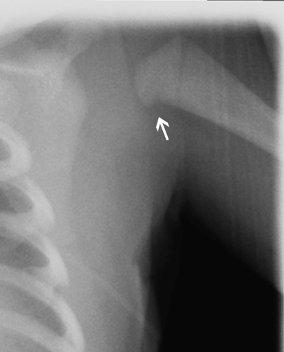
Results: Injuries that are highly specific for the diagnosis of abuse include metaphyseal corner fractures, posteromedial rib fractures, and sternal, scapular, and spinous process fractures. Lesions of moderate specificity include, among other injuries, multiple fractures of various ages and epiphyseal separations. Long-bone fractures and clavicular fractures, while common, are of low specificity. In addition to the appropriate accurate diagnosis of these injuries, several diseases and syndromes may mimic abuse due to the similarity in the radiographic picture.

Conclusions: Stratification of fractures sustained in child abuse according to specificity and an understanding of the several diseases that mimic abuse are helpful in the accurate diagnosis of child abuse.

Level of evidence: Level V, diagnostic study. See Guidelines for Authors for a complete description of levels of evidence.


Figures

Fig. 1A–B
Fig. 1A–B
(A) A frontal chest radiograph of a 2-month-old infant shows a questionable area of rib expansion (arrowhead), not well seen against the background of lung markings. (B) In an oblique view of the same infant, focal rib expansion is more clearly seen, consistent with a healing rib fracture (arrowhead). The linear fracture line is also apparent.
Fig. 2A–B
Fig. 2A–B
(A) An AP chest radiograph of a 3-month-old infant shows a classic posteromedial rib fracture near the midline (arrow), with other healing rib fractures seen elsewhere (arrowheads). (B) An axial CT image of the same patient shows the same posteromedial fracture with irregular linear lucency (arrow). It is more clearly evident on the CT image than on the radiograph shown in (A). Other rib fractures with periosteal reaction indicating healing are also present (arrowheads and curved arrow).
Fig. 3
Fig. 3
An AP chest radiograph of a 1-month-old infant shows multiple right lateral rib fractures. Focal expansion and medullary lucency are present at the eighth rib fracture (arrow) typical of the “hole in the rib” appearance.
Fig. 4
Fig. 4
A high-detail oblique view of the ribs of a 6-month-old infant shows multiple healing posteromedial rib fractures (arrowheads). The level of detail in this image is far greater than what would be present on a standard chest radiograph.
Fig. 5
Fig. 5
An AP radiograph of the knee of a 4-month-old infant shows a CML at the lateral aspect of the distal femoral metaphysis (arrowhead). Note periosteal reaction (curved arrow) seen more proximally and ending at the level of the physis. Also note a healing corner fracture of the proximal tibia (arrow).
Fig. 6
Fig. 6
An AP radiograph of the femur of a 12-day-old infant shows exuberant callus about the distal femoral bucket handle CML. This infant was a difficult footling extraction. Callus is in an advanced stage due to the young age of the patient. This amount of callus would not be present in a more recent fracture.
Fig. 7A–B
Fig. 7A–B
(A) A lateral radiograph of the elbow of a 1-year-old child shows the capitellum (arrow) is posteriorly displaced along with the radius and ulna. The fracture plane has run through the zone of provisional calcification (arrowhead) of the distal humerus. (B) A coronal sonographic image of the distal humerus identifies the fractured and displaced zone of provisional calcification as an irregular curvilinear echogenic density (arrowheads).
Fig. 8
Fig. 8
An AP radiograph of the elbow of a 2-year-old child shows a healing supracondylar fracture (not well seen). Note how the periosteal reaction (arrowheads) ends at the capsular origin (arrow).
Fig. 9
Fig. 9
An AP radiograph of the wrist of a 2-year-old boy shows a focal straightened metaphyseal collar that interrupts the smooth flaring of the metaphysis (arrows).
Fig. 10A–B
Fig. 10A–B
(A) An AP radiograph of the knee of a 3-month-old infant shows a normal metaphyseal spur with the metaphyseal collar extending along the physis (arrowhead). Note the spur is not being undercut by linear lucency and it continues smoothly onto the metaphyses. (B) An AP radiograph of the knee of a 2-month-old infant shows a CML at the distal femur. Note the excrescence medial to the physis (arrow) is not smooth and linear lucency separates it from the edge of the metaphysis. A corner fracture seen more as a bucket handle-type injury is less well seen at the proximal tibia (arrowhead).
Fig. 11
Fig. 11
A lateral view of the knee of a 15-month-old child shows the triangular-shaped fragmented appearance of the posterior aspect of the femur (arrow).
Fig. 12
Fig. 12
A magnified frontal view of the proximal humerus of a 1-month-old infant shows the concave medial surface of the proximal humerus ends in a small beak. A normal notch is seen along the surface (arrow) and is a normal finding at this cutaway zone.
Fig. 13A–B
Fig. 13A–B
(A) A lateral radiograph of the skull of a 3-month-old infant shows a thin linear lucency (arrowhead) at the posterior parietal bone. (B) A 3D reconstruction of the skull in the same patient identifies the linear lucency as a vascular groove along the surface of the skull (arrowheads). This was confirmed using electronic crossreferencing.

Similar articles

Cited by

References

    1. Ablin DS, Sane SM. Non-accidental injury: confusion with temporary brittle bone disease and mild osteogenesis imperfecta. Pediatr Radiol. 1997;27:111–113. doi: 10.1007/s002470050079. - DOI - PubMed
    1. Adams PC, Strand RD, Bresnan MJ, Lucky AW. Kinky hair syndrome: serial study of radiological findings with emphasis on the similarity to the battered child syndrome. Radiology. 1974;112:401–407. - PubMed
    1. American College of Radiology. ACR practice guideline for skeletal surveys in children. Available at: http://www.acr.org/SecondaryMainMenuCategories/quality_safety/guidelines.... Accessed September 11, 2009.
    1. Astley R. Metaphyseal fractures in osteogenesis imperfecta. Br J Radiol. 1979;52:441–443. doi: 10.1259/0007-1285-52-618-441. - DOI - PubMed
    1. Baldridge D, Schwarze U, Morello R, Lennington J, Bertin TK, Pace JM, Pepin MG, Weis M, Eyre DR, Walsh J, Lambert D, Green A, Robinson H, Michelson M, Houge G, Lindman C, Martin J, Ward J, Lemyre E, Mitchell JJ, Krakow D, Rimoin DL, Cohn DH, Byers PH, Lee B. CRTAP and LEPRE1 mutations in recessive osteogenesis imperfecta. Hum Mutat. 2008;29:1435–1442. doi: 10.1002/humu.20799. - DOI - PMC - PubMed