. 2010 Aug 1;110(5):1073-81.
doi: 10.1002/jcb.22619.

Effect of hyperthermia in combination with TRAIL on the JNK-Bim signal transduction pathway and growth of xenograft tumors

Affiliations

Effect of hyperthermia in combination with TRAIL on the JNK-Bim signal transduction pathway and growth of xenograft tumors

Marco A Alcala Jr et al. J Cell Biochem. .

Abstract

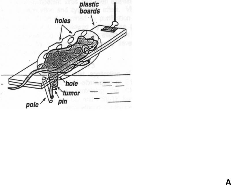
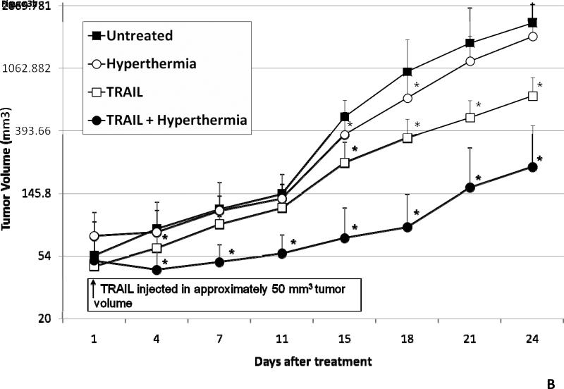
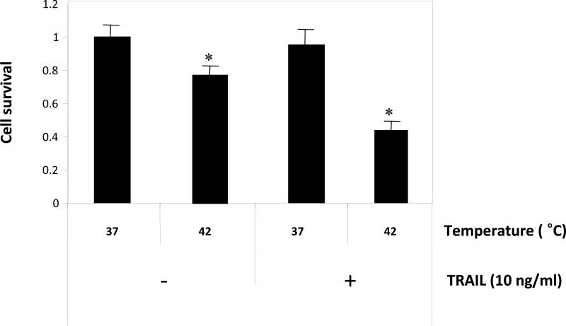
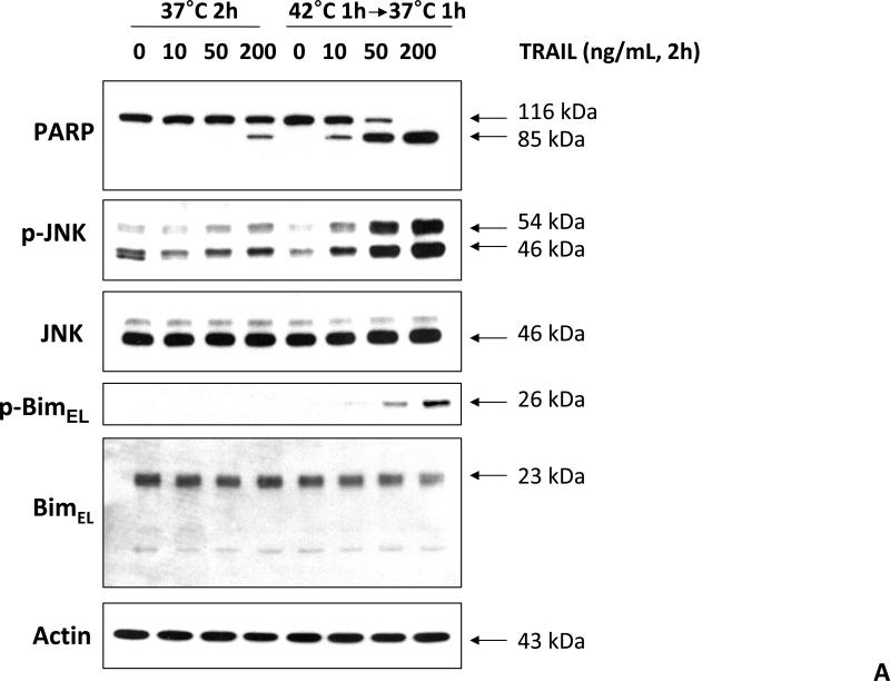
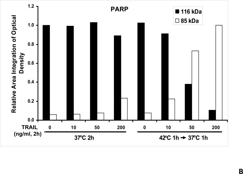
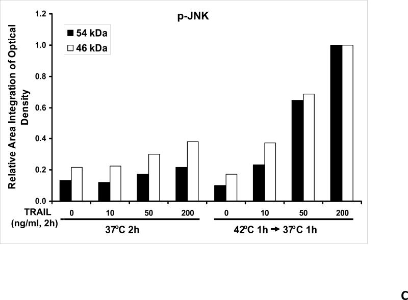
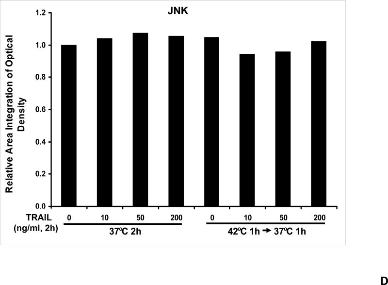
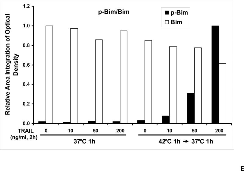
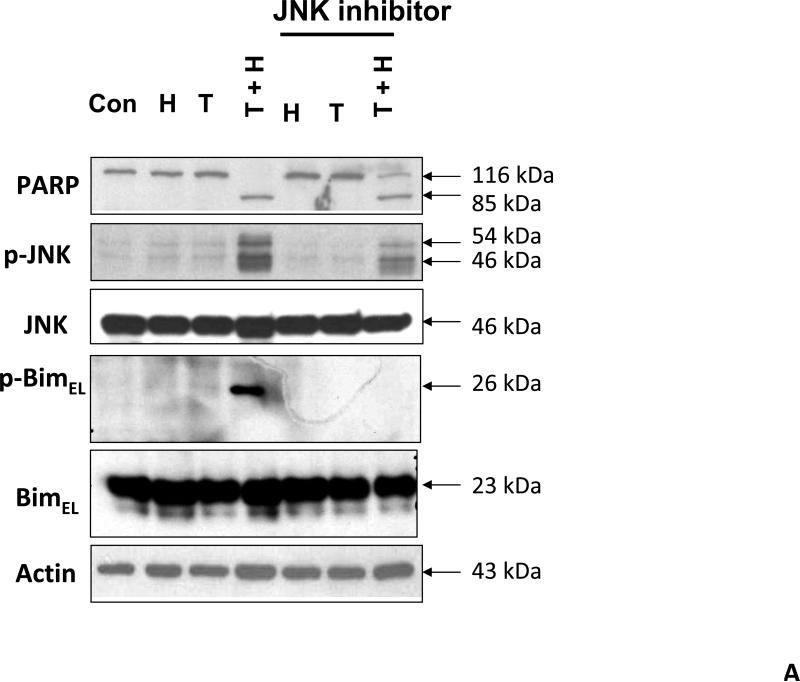
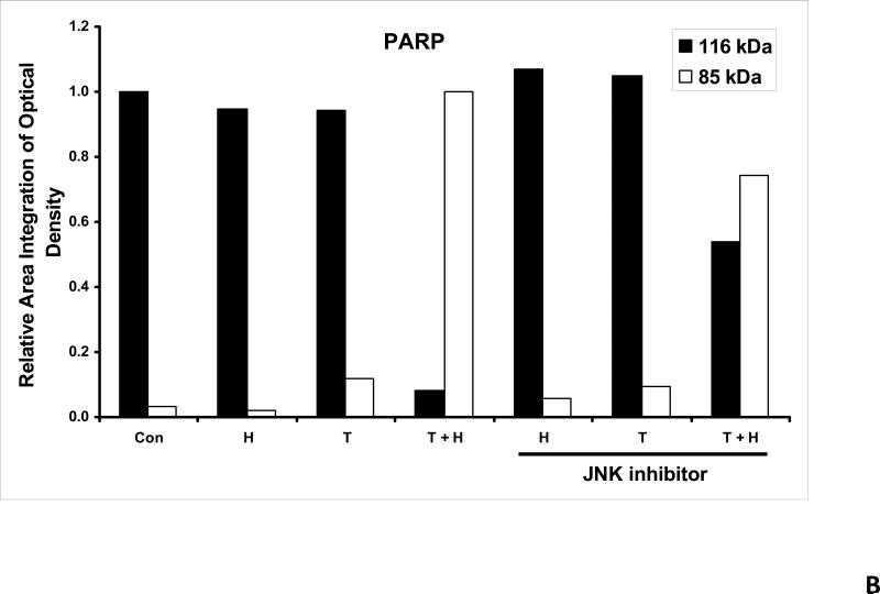
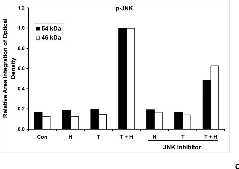
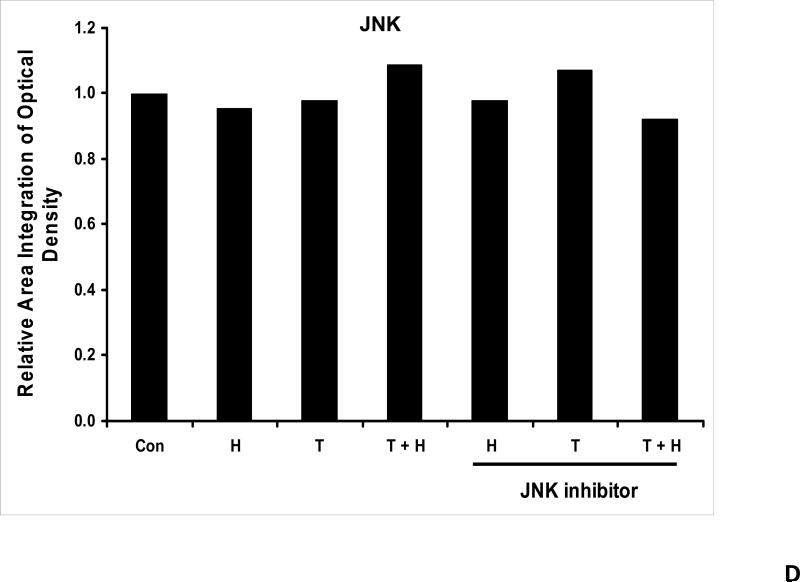
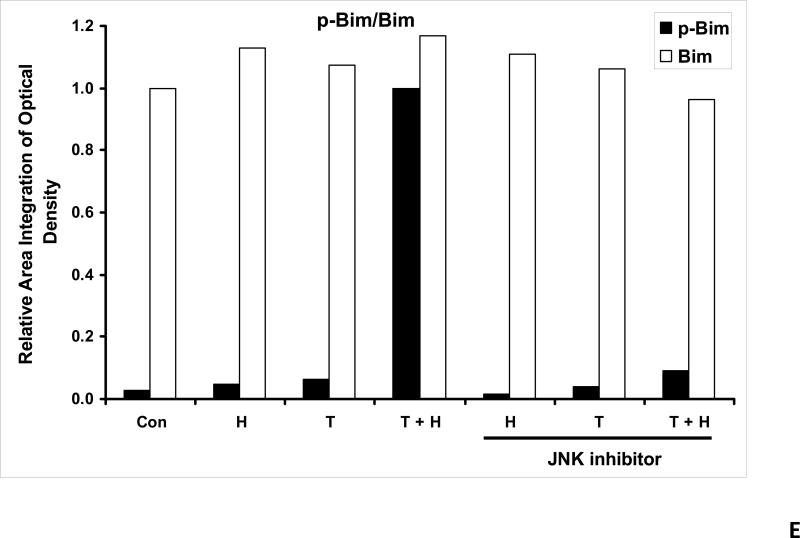
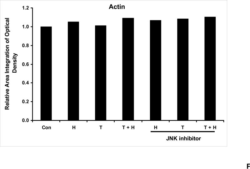
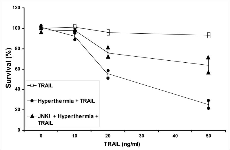
Approximately 25% of patients with colorectal cancer develop metastases to the liver, and surgery is currently the best treatment available. But there are several patients who are unresectable, and isolated hepatic perfusion (IHP) offers a different approach in helping to treat these patients. IHP is a method used for isolating the liver and delivering high doses of chemotherapeutic agents. The efficacy of IHP has been improved by combining hyperthermia not only with chemotherapeutics but with other deliverable agents such as tumor necrosis factor-related apoptosis-inducing ligand (TRAIL). In this study, we used human colorectal cancer CX-1 cells and treated them with hyperthermia and TRAIL, causing cytotoxicity. We were able to demonstrate that the numbers of live cells were significantly reduced with hyperthermia and 10 ng/ml of TRAIL combined. We also showed that the effect of hyperthermia on TRAIL in our studies was enhancement of the apoptotic pathway by the promotion of JNK and Bim(EL) activity as well as PARP cleavage. We have also used our CX-1 cells to generate tumors in Balb/c nude mice. With intratumoral injections of TRAIL combined with hyperthermia at 42 degrees C, we were able to show a delayed onset of tumor growth in our xenograft model.


Figures

Figure 1
Figure 1. Schema of experimental model of heat treatment (A) and effect of hyperthermia, TRAIL, or hyperthermia in combination with TRAIL on growth of CX-1 xenograft (B)
(A) Heating of tumors at a mild hyperthermic temperature (42°C) for 1 hr was performed using water bath immersion of the tumor-bearing right leg. (B) TRAIL was injected into the subcutaneous tumors of mice once they were approximately 50 mm3 in tumor volume. Tumors were exposed to hyperthermic conditions as well as treated with and without TRAIL. Tumors were measured every three days. Error bars represent standard error of the mean from five mice. Asterisk * represents a statistically significant difference between control and hyperthermia alone, TRAIL alone, or hyperthermia in combination with TRAIL at P<0.05.
Figure 2
Figure 2. Effect of hyperthermia in combination with TRAIL on cell viability
CX-1 tumor cells were exposed to hyperthermic (42°C) or normothermic (37°C) conditions for 1 h in conjunction with and without 10 ng/ml of TRAIL. After treatment, cells were then exposed to 37°C for 3 h in the presence of 10 ng/ml of TRAIL. After 3 h exposure, cell survival was determined by colony formation. For colony formation, cells were trypsinized, counted, and plated. After 10 days, colonies were counted and survival was determined as described in Materials and Methods. Error bars represent standard error from the mean (S.E.M.) for three separate experiments. Asterisk * represents a statistically significant difference between control (37°C only) and heat (42°C only) or heat + TRAIL-treated cells at P<0.01.
Figure 3
Figure 3. Effect of hyperthermia in combination with TRAIL on the JNK-Bim signal transduction pathway
(A) CX-1 cells were exposed to hyperthermic conditions (42°C) for 1 h in the presence of TRAIL (10-200 ng/ml) and then incubated for 1 h at 37°C in the presence of TRAIL. Equal amounts of protein (20 μg) from cell lysates were separated by SDS-PAGE and immunoblotted with anti-PARP, anti-phosphorylated JNK, anti-JNK, anti-phospho-Bim, or anti-Bim antibody. Actin was used to confirm the equal amount of proteins loaded in each lane. Hyperthermia combined with moderate to high doses of TRAIL (50 – 200ng/ml) shows an increase in activation of the JNK-Bim signaling transduction pathway demonstrating apoptotic cell death. (B, C, D, E, F) Densitometry analysis of the bands from the JNK-Bim signal pathway for PARP (B), p-JNK (C), JNK (D), pBim/Bim (E), and actin (F) was performed.
Figure 4
Figure 4. Effect of JNK inhibitor on hyperthermia in combination with TRAIL-activated JNK-Bim signal and apoptosis
(A) JNK inhibitor II (50 μg/ml) was added to CX-1 cell cultures 1 h prior to exposing them to hyperthermic (42°C) conditions for 1 h in the presence of TRAIL (50 ng/ml) and then incubated for 1 h at 37°C in the presence of TRAIL. Immunoblotting assay was performed as described in Figure 3A. JNK inhibitor effectively inhibited the JNK-Bim signal and protected cells from apoptosis during treatment with hyperthermia in combination with TRAIL. (B, C, D, E, F) Densitometry analysis of the bands from the JNK-Bim signal pathway with/without JNK inhibitor for PARP (B), p-JNK (C), JNK (D), pBim/Bim (E), and actin (F) was performed.
Figure 5
Figure 5. Effect of JNK inhibitor (JNKI) on hyperthermia in combination with TRAIL-induced cytotoxicity in CX-1 cells
JNK inhibitor II (50 μg/ml) was added for 1 h prior to the treatment of hyperthermia (42°C) for 1 h in the presence of TRAIL (10 – 50 ng/ml). After treatment, cells were exposed to 37°C for 1 h in the presence of TRAIL (10 – 50 ng/ml). Cell survival was immediately determined by the trypan blue exclusion assay as described by Materials and Methods. Duplicate experiments were performed.

References

    1. Ashkenazi A, Pai RC, Fong S, Leung S, Lawrence DA, Marsters SA, Blackie C, Chang L, McMurtrey AE, Hebert A, DeForge L, Koumenis IL, Lewis D, Harris L, Bussiere J, Koeppen H, Shahrokh Z, Schwall RH. Safety and antitumor activity of recombinant soluble Apo2 ligand. J Clin Invest. 1999;104:155–162. - PMC - PubMed
    1. Bartlett DL, Libutti SK, Figg WD, Fraker DL, Alexander HR. Isolated hepatic perfusion for unresectable hepatic metastases from colorectal cancer. Surgery. 2001;129:176–187. - PubMed
    1. Boveris A, Cadenas E. Production of superoxide radicals and hydrogen peroxide in mitochondria. In: Oberley LW, editor. Superoxide Dismutase. II. CRC Press Inc.; Boca Raton, FL: 1982. pp. 15–30.
    1. Chen YR, Wang W, Kong AN, Tan TH. Molecular mechanisms of c-Jun N-terminal kinase-mediated apoptosis induced by anticarcinogenic isothiocyanates. J Biol Chem. 1998;2733:1769–1775. - PubMed
    1. Erkmen K, Egorin MJ, Reyno LM, Morgan R, Jr., Doroshow JH. Effects of storage on the binding of carboplatin to plasma proteins. Cancer Chemother Pharmacol. 1995;35:254–256. - PubMed

Publication types

MeSH terms