Review
. 2010 Nov 1;88(14):2991-3001.
doi: 10.1002/jnr.22444.

The decade of the dendritic NMDA spike

Affiliations
Review

The decade of the dendritic NMDA spike

Srdjan D Antic et al. J Neurosci Res. .

Abstract

In the field of cortical cellular physiology, much effort has been invested in understanding thick apical dendrites of pyramidal neurons and the regenerative sodium and calcium spikes that take place in the apical trunk. Here we focus on thin dendrites of pyramidal cells (basal, oblique, and tuft dendrites), and we discuss one relatively novel form of an electrical signal ("NMDA spike") that is specific for these branches. Basal, oblique, and apical tuft dendrites receive a high density of glutamatergic synaptic contacts. Synchronous activation of 10-50 neighboring glutamatergic synapses triggers a local dendritic regenerative potential, NMDA spike/plateau, which is characterized by significant local amplitude (40-50 mV) and an extraordinary duration (up to several hundred milliseconds). The NMDA plateau potential, when it is initiated in an apical tuft dendrite, is able to maintain a good portion of that tuft in a sustained depolarized state. However, if NMDA-dominated plateau potentials originate in proximal segments of basal dendrites, they regularly bring the neuronal cell body into a sustained depolarized state, which resembles a cortical Up state. At each dendritic initiation site (basal, oblique, and tuft) an NMDA spike creates favorable conditions for causal interactions of active synaptic inputs, including the spatial or temporal binding of information, as well as processes of short-term and long-term synaptic modifications (e.g., long-term potentiation or long-term depression). Because of their strong amplitudes and durations, local dendritic NMDA spikes make up the cellular substrate for multisite independent subunit computations that enrich the computational power and repertoire of cortical pyramidal cells. We propose that NMDA spikes are likely to play significant roles in cortical information processing in awake animals (spatiotemporal binding, working memory) and during slow-wave sleep (neuronal Up states, consolidation of memories).


Figures

Figure 1
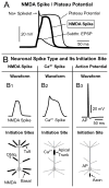
Figure 1. Each Class of Neurites (Basal, Oblique, Tuft, Apical Trunk, and Axon) Supports a Characteristic Regenerative Potential (Spike)
(A) In thin dendrites of pyramidal neurons (basal, oblique and tuft) weak glutamatergic inputs produce EPSP-like depolarizations (“Subthreshold EPSP”). Stronger (suprathreshold) inputs regularly trigger dendritic “Plateau Potentials”. Plateau potentials are characterized by a rapid onset, initial spikelet (Na+ Spikelet), plateau phase, and an abrupt collapse at the end of the plateau phase. A glutamate-evoked plateau potential is the product of several dendritic conductances. The major ionic contributor is regenerative NMDA receptor current. Block of sodium and calcium channels with TTX and Cd2+, respectively, reveals a pure “NMDA Spike” in the dendrite (Schiller et al., 2000). (B) The waveforms of three regenerative potentials (spikes) are shown side by side on the same scale (B1-B3). The neuronal compartment which serves as the spike initiation site is colored black below each spike waveform (Initiation Sites). The principle compartments of cortical pyramidal cells are: Soma, Axon, Apical Trunk, Basal, Oblique and Tuft dendrites. (B1) NMDA spikes last approx-imately 50–100 ms and they can be initiated in the apical tuft, apical oblique and basal dendrites. (B2) Calcium spikes have slightly greater amplitudes and durations than NMDA spikes. Calcium spikes are often found in the calcium spike initiation zone on the apical trunk. (B3) Action potential (AP) initiates in the axon initial segment. Once initiated in the axon the AP propagates back into the dendritic tree (Stuart et al., 1997). The voltage waveforms of a backpropagating action potential at dendritic sites are not shown in this figure (see (Antic, 2003; Nevian et al., 2007; Zhou et al., 2008; Holthoff et al., 2010)). Dendritic-spike initiation sites (B1-B3) are based on Larkum et al., 2009.
Figure 2
Figure 2. Strict correlation between dendritic plateau potential and somatic sustained depolarization
(A) Recording configuration showing glutamate iontophoresis electrode positioned in the middle segment of a basal dendrite. (B) Voltage waveforms are recorded at the stimulation site optically (dend.) and in the cell body electrically (patch) as indicated in “A”. Intensity of glutamate current pulse was increased in 4 equal increments (1 – 4). In this and the following panels the timing of glutamate pulse is marked by a vertical tick below the recording trace. In the presence of TTX the somatic membrane potential waveform closely follows that in the dendrite. While the amplitudes of both dendritic and somatic responses are saturated, their durations increase gradually in consecutive sweeps. There is a positive correlation between the stimulus intensity and duration of the electrical event (Milojkovic et al., 2005a). (C) In normal physiological saline the glutamate-evoked dendritic plateau potential is often preceded by a fast sodium spikelet. At this same moment when the spikelet fired in the dendrite the cell body showed fast inflection (arrow)(Milojkovic et al., 2005b; Nevian et al., 2007; Remy et al., 2009). (D) Somatic voltage waveforms generated by a sequence of incrementally increasing glutamate stimuli delivered on a basal dendrite, as shown in “A”. (E) Slow component of the somatic depolarization can be described in terms of amplitude (amp.) and half-width (duration, dur.). The amplitude and duration of the slow component (somatic) are plotted against stimulus intensity in F and G, respectively. (F) The amplitude of somatic slow depolarization exhibits an all-or-none highly non-linear behavior (Binary), while duration exhibits steady growth with stimulus intensity (Linear “G”). (H) Cortical UP state-like somatic depolarizations were triggered in acute brain slice by a sequence of three identical glutamate puffs (glut.) delivered on a single basilar branch (Milojkovic et al., 2004).
Figure 3
Figure 3. Two-Directional Propagation of Glutamate-evoked Dendritic Plateau Potentials
The dendritic plateau potential (Plateau Potential) propagates from its initiation site (Glut. Input) in two directions: Distally towards the end of the basal dendrite (Dend. Tip); and proximally towards the cell body (Soma). In the proximal direction (white arrows) the rate of amplitude decline is much faster than in the distal direction (grey arrows). Glutamate-evoked dendritic plateau potentials initiated in the mid section of a typical basal dendrite (rectangle) regularly cause 10 – 20 mV sustained (> 100 ms) depolarizations of the cell body (Milojkovic et al., 2004; Milojkovic et al., 2005a). Note that at the glutamate stimulation site (Glut. Input) the dendritic plateau potential is followed by a small post-plateau depolarization (Post-plateau Potential). Both, the plateau and post-plateau potential, support calcium influxes, which when combined constitute the glutamate-evoked dendritic “calcium plateau”, not shown, but see (Milojkovic et al., 2007).

Similar articles

Cited by

References

    1. Acker CD, Antic SD. Quantitative assessment of the distributions of membrane conductances involved in action potential backpropagation along Basal dendrites. J Neurophysiol. 2009;101:1524–1541. - PMC - PubMed
    1. Antic SD. Action potentials in basal and oblique dendrites of rat neocortical pyramidal neurons. J Physiol. 2003;550:35–50. - PMC - PubMed
    1. Antic SD, Acker CD, Zhou WL, Moore AR, Milojkovic BA. The Role of Dendrites in the Maintenance of the UP State. In: Timofeev I, editor. Mechanisms of spontaneous active states in the neocortex, Mechanisms of spontaneous active states in the neocortex Edition. Research Signpost; Kerala, India: 2007. pp. 45–72.
    1. Archie KA, Mel BW. A model for intradendritic computation of binocular disparity. Nature Neuroscience. 2000;3:54–63. - PubMed
    1. Ariav G, Polsky A, Schiller J. Submillisecond precision of the input-output transformation function mediated by fast sodium dendritic spikes in basal dendrites of CA1 pyramidal neurons. J Neurosci. 2003;23:7750–7758. - PMC - PubMed

Publication types

Substances