

The mitochondrial proteome: a dynamic functional program in tissues and disease states
- PMID: 20544878
- PMCID: PMC3209511
- DOI: 10.1002/em.20574
The mitochondrial proteome: a dynamic functional program in tissues and disease states
Abstract
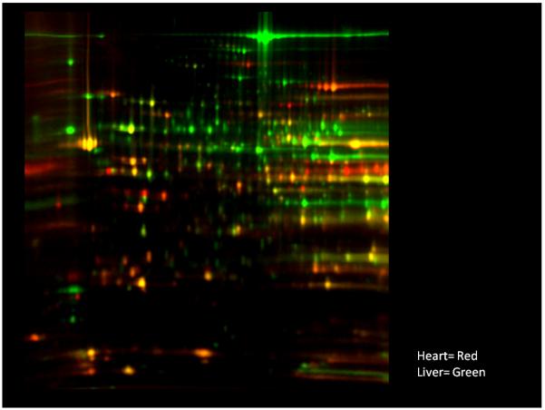
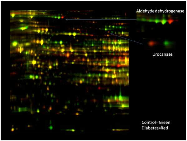
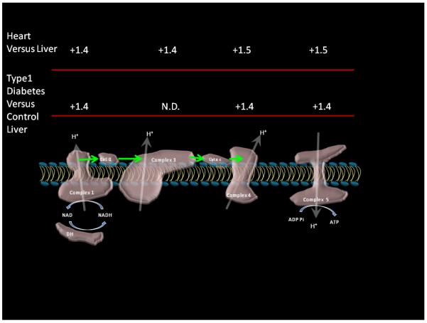
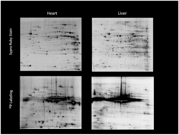
The nuclear DNA transcriptional programming of the mitochondria proteome varies dramatically between tissues depending on its functional requirements. This programming generally regulates all of the proteins associated with a metabolic or biosynthetic pathway associated with a given function, essentially regulating the maximum rate of the pathway while keeping the enzymes at the same molar ratio. This may permit the same regulatory mechanisms to function at low- and high-flux capacity situations. This alteration in total protein content results in rather dramatic changes in the mitochondria proteome between tissues. A tissues mitochondria proteome also changes with disease state, in Type 1 diabetes the liver mitochondrial proteome shifts to support ATP production, urea synthesis, and fatty acid oxidation. Acute flux regulation is modulated by numerous posttranslational events that also are highly variable between tissues. The most studied posttranslational modification is protein phosphorylation, which is found all of the complexes of oxidative phosphorylation and most of the major metabolic pathways. The functional significance of these modifications is currently a major area of research along with the kinase and phosphatase regulatory network. This near ubiquitous presence of protein phosphorylations, and other posttranslational events, in the matrix suggest that not all posttranslational events have functional significance. Screening methods are being introduced to detect the active or dynamic posttranslational sites to focus attention on sites that might provide insight into regulatory mechanisms.
Figures
References
-
- Aden N, Shiwen X, Aden D, Black C, Nuttall A, Denton CP, et al. Proteomic analysis of scleroderma lesional skin reveals activated wound healing phenotype of epidermal cell layer. Rheumatology. 2008;47:1754–1760. - PubMed
-
- Aitken RJ, Baker MA. The role of proteomics in understanding sperm cell biology. Int J Andrology. 2007;31:295–302. - PubMed
-
- Anello M, Spampinato D, Piro S, Purrello F, Rabuazzo AM. Glucosamine-induced alterations of mitochondrial function in pancreatic beta-cells: possible role of protein glycosylation. Am J Physiol Endocrinol Metab. 2004;287:E602–E608. - PubMed
Publication types
MeSH terms
Substances
Grants and funding
LinkOut - more resources
Full Text Sources
