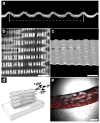
Microcrimped collagen fiber-elastin composites
- PMID: 20544890
- PMCID: PMC3213053
- DOI: 10.1002/adma.200903612
Microcrimped collagen fiber-elastin composites
Figures


Similar articles
-
Preparation and characterization of novel elastin-like polypeptide-collagen composites.J Biomed Mater Res A. 2013 Aug;101(8):2383-91. doi: 10.1002/jbm.a.34514. Epub 2013 Feb 20. J Biomed Mater Res A. 2013. PMID: 23427027
-
The use of microfiber composites of elastin-like protein matrix reinforced with synthetic collagen in the design of vascular grafts.Biomaterials. 2010 Sep;31(27):7175-82. doi: 10.1016/j.biomaterials.2010.05.014. Epub 2010 Jun 26. Biomaterials. 2010. PMID: 20584549 Free PMC article.
-
Generation of spatially aligned collagen fiber networks through microtransfer molding.Adv Healthc Mater. 2014 Mar;3(3):367-74. doi: 10.1002/adhm.201300112. Epub 2013 Aug 29. Adv Healthc Mater. 2014. PMID: 24039146 Free PMC article.
-
Elastin-based materials.Chem Soc Rev. 2010 Sep;39(9):3371-9. doi: 10.1039/b919452p. Epub 2010 May 7. Chem Soc Rev. 2010. PMID: 20449520 Review.
-
Recombinant elastin-mimetic biomaterials: Emerging applications in medicine.Adv Drug Deliv Rev. 2010 Dec 30;62(15):1468-78. doi: 10.1016/j.addr.2010.04.007. Epub 2010 May 2. Adv Drug Deliv Rev. 2010. PMID: 20441783 Free PMC article. Review.
Cited by
-
Spontaneous Crimping of Gelatin Methacryloyl Nanofibrils Induced by Limited Hydration.ACS Biomater Sci Eng. 2025 Aug 11;11(8):4758-4772. doi: 10.1021/acsbiomaterials.5c00828. Epub 2025 Jul 18. ACS Biomater Sci Eng. 2025. PMID: 40679322 Free PMC article.
-
From fiber curls to mesh waves: a platform for the fabrication of hierarchically structured nanofibers mimicking natural tissue formation.Nanoscale. 2019 Aug 1;11(30):14312-14321. doi: 10.1039/c8nr10108f. Nanoscale. 2019. PMID: 31322143 Free PMC article.
-
Challenges in Translating from Bench to Bed-Side: Pro-Angiogenic Peptides for Ischemia Treatment.Molecules. 2019 Mar 28;24(7):1219. doi: 10.3390/molecules24071219. Molecules. 2019. PMID: 30925755 Free PMC article. Review.
-
Extracellular matrix-based intracortical microelectrodes: Toward a microfabricated neural interface based on natural materials.Microsyst Nanoeng. 2015;1(1):15010. doi: 10.1038/micronano.2015.10. Epub 2015 Jun 29. Microsyst Nanoeng. 2015. PMID: 30498620 Free PMC article.
-
The Role of the Non-Collagenous Extracellular Matrix in Tendon and Ligament Mechanical Behavior: A Review.J Biomech Eng. 2022 May 1;144(5):050801. doi: 10.1115/1.4053086. J Biomech Eng. 2022. PMID: 34802057 Free PMC article. Review.
References
Publication types
MeSH terms
Substances
Grants and funding
LinkOut - more resources
Full Text Sources

