

doi: 10.1002/anie.201000378.
Alteration of the alpha-synuclein folding landscape by a mutation related to Parkinson's disease
Affiliations
- PMID: 20544898
- PMCID: PMC2972640
- DOI: 10.1002/anie.201000378
Item in Clipboard
Alteration of the alpha-synuclein folding landscape by a mutation related to Parkinson's disease
Angew Chem Int Ed Engl.
.
Free PMC article
No abstract available
Figures
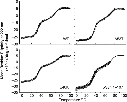
Three-state thermal unfolding of WT α-synuclein, the PD-linked A53T and E46K mutants, and a C-terminal-truncation variant (residues 1–107). Protein-denaturation transitions were monitored by far-UV CD spectroscopy in the presence of SDS (1 mm ). Unfolding data were collected from 0 to 100°C for all peptides except αSyn 1–107 (0–70°C), which aggregates at higher temperatures under the experimental conditions used (see the Supporting Information for additional details).
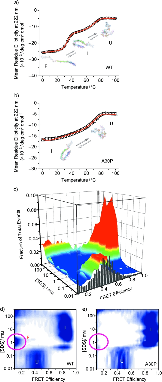
Single-molecule and ensemble characterization of the effect of the PD-linked A30P mutation on the coupled binding and folding of α-synuclein. a,b) Thermal unfolding of WT α-synuclein (a) and the A30P mutant (b) bound to SDS monomers, as monitored at the ensemble level by CD spectroscopy in the presence of SDS (1 mm ). c) Conformational transitions of A30P α-synuclein induced by changes in ligand concentration at room temperature, as detected at the single-molecule level by smFRET. d) Two-dimensional contour plot of previously reported WT data for comparison with the corresponding plot for the A30P mutant in (e). e) Two-dimensional contour plot of the raw data presented in (c). U, I, and F are different protein conformational states that exhibit characteristic FRET efficiency and helicity. U is the natively unfolded state, I corresponds to a bent helical structure previously solved by NMR spectroscopy (1XQ8), and F is an extended α-helix structure. (See the Supporting Information for additional details).
Similar articles
-
Effect of reparation of repeat sequences in the human alpha-synuclein on fibrillation ability.Int J Biol Sci. 2006 Oct 2;3(1):1-7. doi: 10.7150/ijbs.3.1. Int J Biol Sci. 2006. PMID: 17200685 Free PMC article.
-
Parkinson's disease as a member of Prion-like disorders.Virus Res. 2015 Sep 2;207:38-46. doi: 10.1016/j.virusres.2014.10.016. Epub 2014 Nov 4. Virus Res. 2015. PMID: 25456401 Review.
-
The newly discovered Parkinson's disease associated Finnish mutation (A53E) attenuates α-synuclein aggregation and membrane binding.Biochemistry. 2014 Oct 21;53(41):6419-21. doi: 10.1021/bi5010365. Epub 2014 Oct 10. Biochemistry. 2014. PMID: 25268550
-
Methamphetamine binds to α-synuclein and causes a conformational change which can be detected by nanopore analysis.FEBS Lett. 2012 Sep 21;586(19):3222-8. doi: 10.1016/j.febslet.2012.06.040. Epub 2012 Jul 4. FEBS Lett. 2012. PMID: 22771474
-
DNA induced folding/fibrillation of alpha-synuclein: new insights in Parkinson's disease.Front Biosci (Landmark Ed). 2010 Jan 1;15(2):418-36. doi: 10.2741/3628. Front Biosci (Landmark Ed). 2010. PMID: 20036828 Review.
Cited by
-
Function and dysfunction of α-synuclein: probing conformational changes and aggregation by single molecule fluorescence.Mol Neurobiol. 2013 Apr;47(2):622-31. doi: 10.1007/s12035-012-8338-x. Epub 2012 Sep 16. Mol Neurobiol. 2013. PMID: 22983916 Free PMC article. Review.
-
Aggregation of α-synuclein is kinetically controlled by intramolecular diffusion.Proc Natl Acad Sci U S A. 2012 Feb 14;109(7):2336-41. doi: 10.1073/pnas.1109526109. Epub 2012 Jan 27. Proc Natl Acad Sci U S A. 2012. PMID: 22308332 Free PMC article.
-
Probing the Basis of α-Synuclein Aggregation by Comparing Simulations to Single-Molecule Experiments.Biophys J. 2019 Sep 17;117(6):1125-1135. doi: 10.1016/j.bpj.2019.08.013. Epub 2019 Aug 16. Biophys J. 2019. PMID: 31477241 Free PMC article.
-
Untangling the Conformational Polymorphism of Disordered Proteins Associated With Neurodegeneration at the Single-Molecule Level.Front Mol Neurosci. 2020 Jan 10;12:309. doi: 10.3389/fnmol.2019.00309. eCollection 2019. Front Mol Neurosci. 2020. PMID: 31998071 Free PMC article. Review.
-
Structures of the E46K mutant-type α-synuclein protein and impact of E46K mutation on the structures of the wild-type α-synuclein protein.ACS Chem Neurosci. 2013 Mar 20;4(3):498-508. doi: 10.1021/cn3002027. Epub 2013 Jan 30. ACS Chem Neurosci. 2013. PMID: 23374074 Free PMC article.
References
Publication types
MeSH terms
Substances
Grants and funding
LinkOut - more resources
Full Text Sources
Medical
