. 2010 Aug;40(8):1203-13.
doi: 10.1111/j.1365-2222.2010.03546.x. Epub 2010 Jun 7.

Human monocytic cells direct the robust release of CXCL10 by bronchial epithelial cells during rhinovirus infection

Affiliations

Human monocytic cells direct the robust release of CXCL10 by bronchial epithelial cells during rhinovirus infection

N L Korpi-Steiner et al. Clin Exp Allergy. 2010 Aug.

Abstract

Background: Human rhinovirus (HRV) infections are a major cause of exacerbations in chronic respiratory conditions such as asthma and chronic obstructive pulmonary disease, but HRV-induced immune responses of the lower airway are poorly understood. Earlier work examining cytokine release following HRV infection has focused on epithelial cells because they serve as the principal site of viral replication, and internalization and replication of viral RNA appear necessary for epithelial cell mediator release. However, during HRV infection, only a small proportion of epithelial cells become infected. As HRV-induced cytokine levels in vivo are markedly elevated, this observation suggests that other mechanisms independent of direct viral infection may induce epithelial cell cytokine release.

Objective: Our aim was to test for the importance of interactions between human bronchial epithelial cells (HBECs) and monocytic cells in the control of mediator release during HRV exposure.

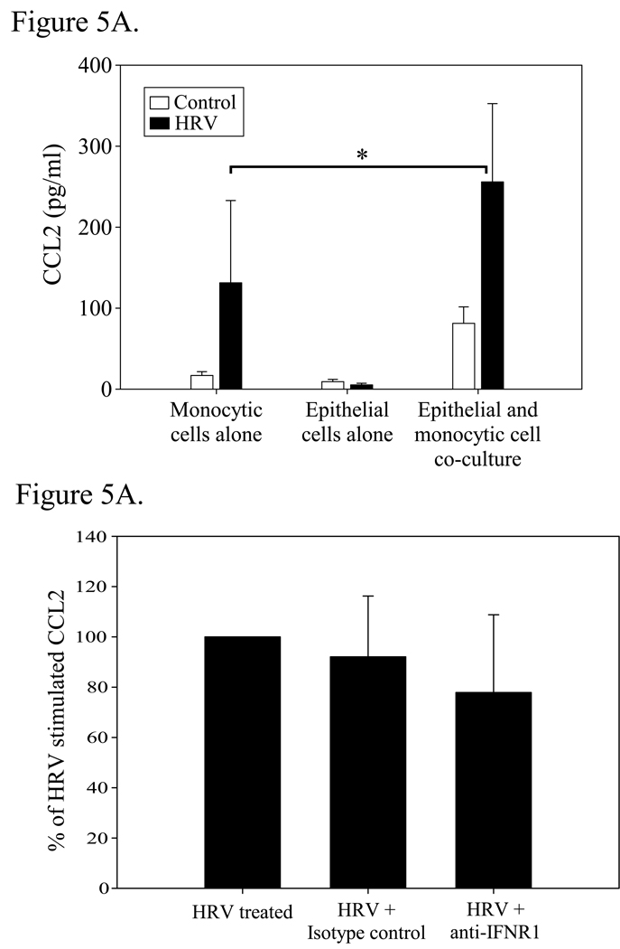
Methods: In vitro models of HRV serotype-16 (HRV16) infection of primary HBECs and human monocytic cells, in mono or co-culture, were used. We assessed HRV16-induced CXCL10 and CCL2 protein release via ELISA.

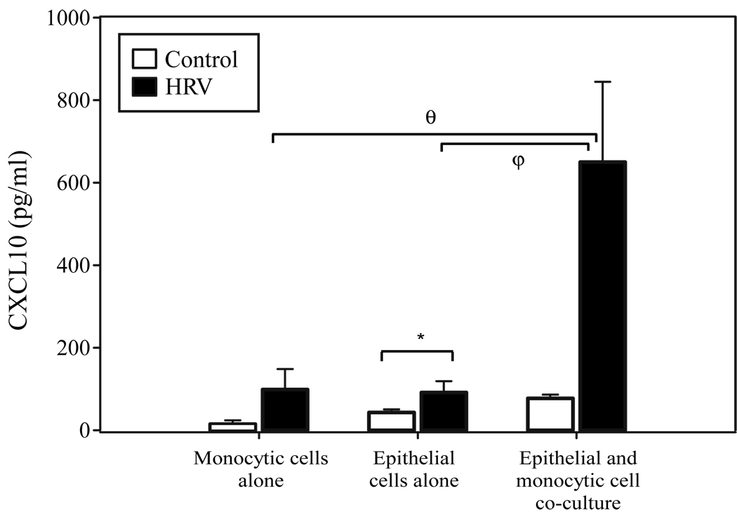
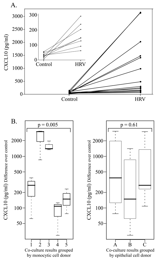
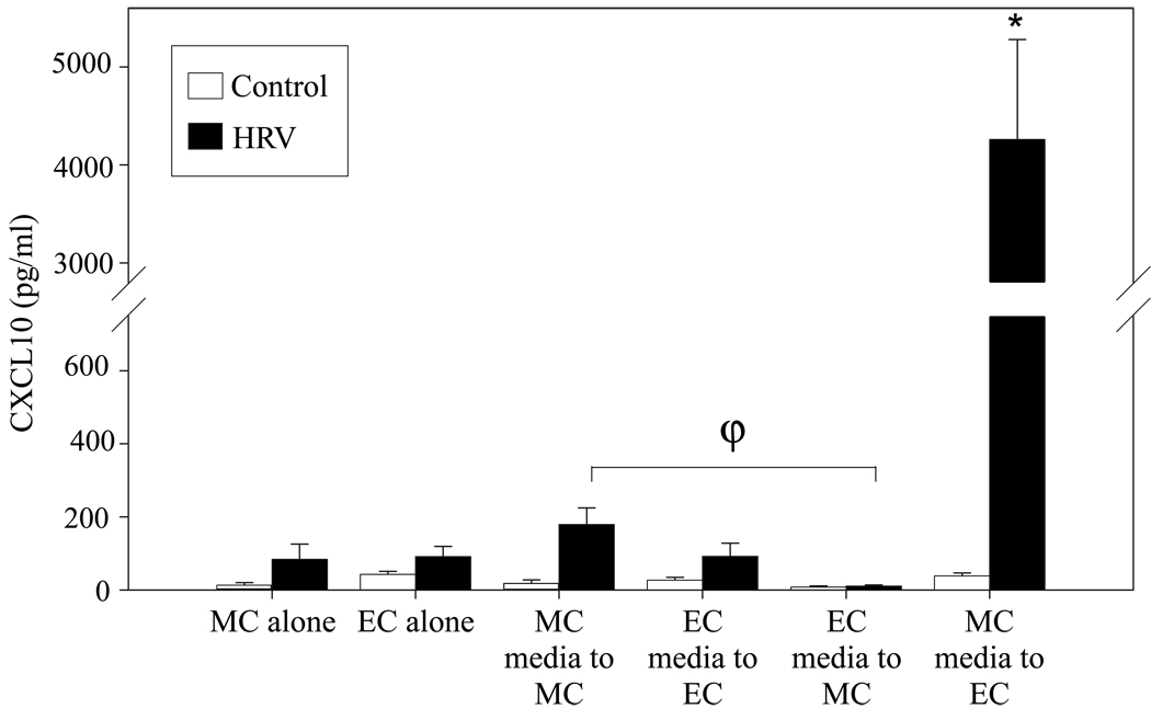
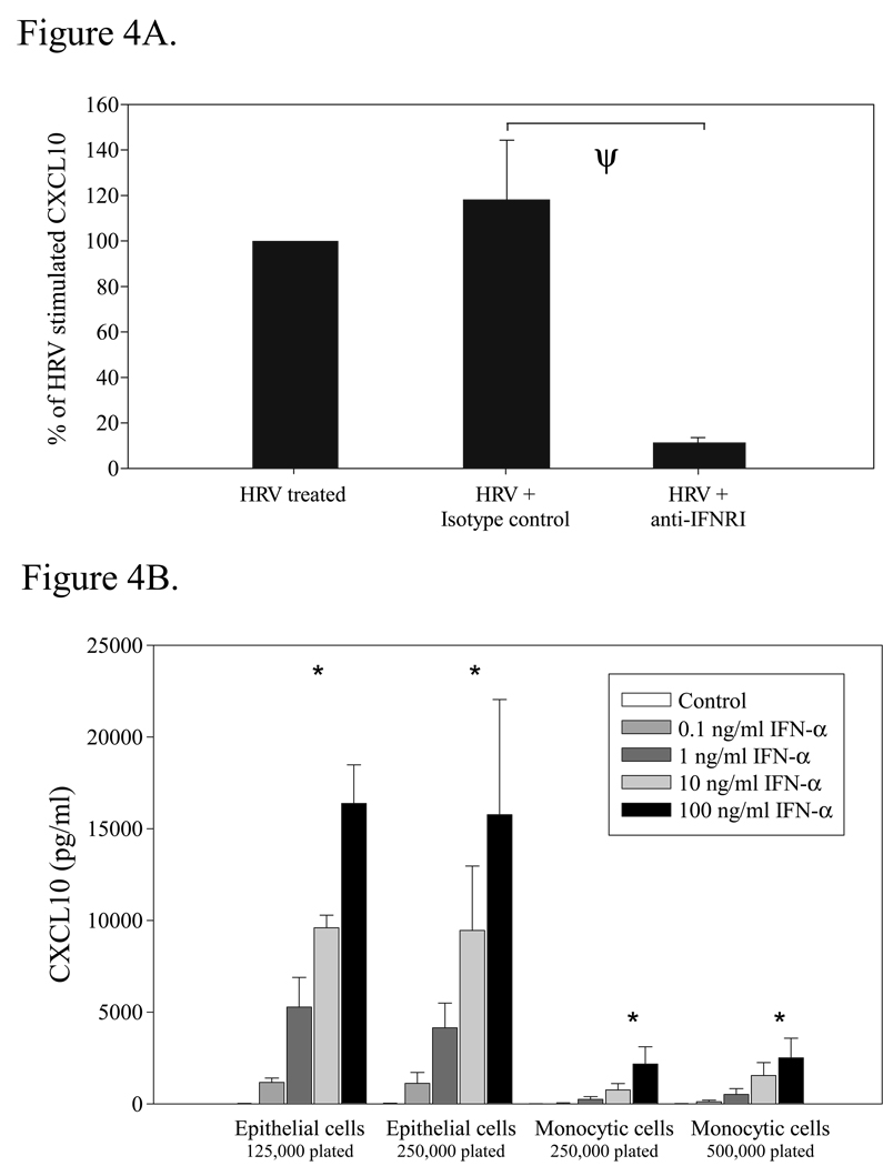
Results: Co-culture of human monocytic and bronchial epithelial cells promoted a synergistic augmentation of CXCL10 and CCL2 protein release following HRV16 challenge. Transfer of conditioned media from HRV16-treated monocytic cells to epithelial cultures induced a robust release of CXCL10 by the epithelial cells. This effect was greatly attenuated by type I IFN receptor blocking antibodies, and could be recapitulated by IFN-alpha addition.

Conclusions: Our data indicate that epithelial CXCL10 release during HRV infection is augmented by a monocytic cell-dependent mechanism involving type I IFN(s). Our findings support a key role for monocytic cells in the amplification of epithelial cell chemokine production during HRV infection, and help to explain how an inflammatory milieu is created in the lower airways even in the absence of extensive viral replication and epithelial infection.


Figures

Figure 1
Figure 1
CXCL10 secretion from HRV16-simulated monocytic cell mono-cultures, epithelial cell mono-cultures, and epithelial-monocytic cell co-cultures. Bronchial epithelial cells were plated at a density of 1.25 × 10⁵ cells/ml (for co-culture) or 2.5 × 10⁵ cells/ml (for mono-culture) in 6-well collagen-coated tissue culture plates. The following day monocytes were plated at a cell density of 5 × 10⁵ cells/ml for mono-cultures and 2.5 × 10⁵ cells/ml for epithelial-monocytic cell co-cultures. Cells were incubated in reduced-hydrocortisone (0.001%HC) BEGM for 24 h prior to stimulation, then stimulated with vehicle control (HBSS + 0.1% BSA) or HRV16 (MOI = 10; resuspended in HBSS + 0.1% BSA) for 48 h (*, p= 0.05; φ, p < 0.03; θ, p < 0.004 by mixed-effects ANOVA). Data are summarized as the mean ± SEM from 6 experiments.
Figure 2
Figure 2
Examination of the variability in HRV-induced CXCL10 release according to cell type in co-culture. Human monocytic cells and bronchial epithelial cells were co-cultured and treated as described in Fig. 1. A. CXCL10 release by epithelial-monocytic cell co-cultures in response to control or HRV16 stimulation. Epithelial cells from 3 different donors and monocytic cells from 5 different donors were used to make 15 co-culture combinations, which showed variable CXCL10 release in response to HRV16. The inset magnifies the lower portion of the graph to show increased CXCL10 release in response to HRV16 for all co-cultures. B. HRV16-induced CXCL10 results from all 15 co-cultures plotted by donor cell type to examine the source of the variability. Control-treated CXCL10 values were subtracted from HRV16-treated for each epithelial/monocytic cell co-culture. On the left, results were plotted according to the monocytic cell donor, with each boxplot indicating CXCL10 release from co-cultures of 3 different EC donors with the same monocytic cell donor. On the right, the same 15 co-culture results were plotted according to the EC donor, with each boxplot indicating CXCL10 release from co-cultures of 5 different monocytic cell donors with the same EC donor.
Figure 3
Figure 3
Effect of cell-conditioned medium on CXCL10 production by epithelial and monocytic cells. Human bronchial epithelial cells and monocytic cells were plated in mono-culture as described in Fig. 1. After treatment with HRV16 (MOI=10) or vehicle control for 24 h, the cell-conditioned media was centrifuged at 500 × g for 5 min at 4°C, and then transferred to mono-cultures of the same or the other cell type. After 48 h, the supernatants were collected for analysis of CXCL10 protein concentration. In parallel, mono-cultures of epithelial cells and monocytic cells were exposed to control or HRV16 for 48 h. Release of CXCL10 protein by epithelial cell cultures that received conditioned medium from HRV16 treated monocytic cells was significantly increased over all other conditions. (*, p<0.015 by mixed-effects ANOVA for all comparisons; ϕ, p<0.03). EC = epithelial cell, MC = monocytic cell. Data represent mean ± SEM from 3 or more experiments.
Figure 4
Figure 4
Role of type I interferons in HRV-induced CXCL10 release by epithelial and monocytic cells. Human bronchial epithelial cells and monocytic cells were plated in co-culture (A) or mono-culture (B) as described in Fig. 1. A. Co-cultures were pretreated with isotype control (IgG2aκ; 5 µg/mol) or anti-IFN receptor type I blocking antibody (anti-IFNRI; 5 µg/mol) for 30 min, then stimulated with vehicle control (HBSS + 0.1% BSA) or HRV16 (MOI = 10) for 48 h. (ψ, p<0.0001 for comparison to HRV + IgG by mixed-effects ANOVA). The data represent the percent of CXCL10 released compared to HRV16-treated cells ± SEM of % treated from 4 independent experiments. B. Epithelial and monocytic cells were plated to reflect the cell numbers present during mono or co-culture experiments, with half the number of each cell type present in co-culture in order to keep total cell number constant. Cells were treated with vehicle control (HBSS + 0.1% BSA) or doses of the type I interferon IFNα2b ranging from 0.1–100 ng/ml (resuspended in HBSS + 0.1% BSA), and then incubated for 48 h at 37°C. The dose response was significant within all conditions (*, p<0.0001 by mixed-effects ANOVA), and between epithelial cell and monocytic cell responses (p<0.0001 by mixed-effects ANOVA). Data represent mean ± SEM from 4 experiments.
Figure 5
Figure 5
Secretion of CCL2 (MCP-1) by epithelial/monocytic cell mono and co-cultures in response to HRV16. A. Bronchial epithelial cells and monocytic cells were plated in mono or co-culture as described in Fig. 1, and treated with vehicle control or HRV16 for 48h. Supernatants were analysed for the presence of CCL2 by ELISA. (*, p=0.05). Data represent mean ± SEM from 3 independent experiments. B. Effect of type I interferon receptor blockade on the release of CCL2. Co-cultures were treated as described in 4A, and then examined for CCL2 levels by ELISA after 48h. Data are expressed as the percent of CCL2 released from HRV16-treated cells ± SEM of % treated from 3 independent experiments.

Similar articles

Cited by

References

    1. Pattemore PK, Johnston SL, Bardin PG. Viruses as precipitants of asthma symptoms. I. Epidemiology. Clin Exp Allergy. 1992;22:325–336. - PMC - PubMed
    1. Nicholson KG, Kent J, Ireland DC. Respiratory viruses and exacerbations of asthma in adults. Bmj. 1993;307:982–986. - PMC - PubMed
    1. Johnston SL, Pattemore PK, Sanderson G, et al. Community study of role of viral infections in exacerbations of asthma in 9–11 year old children. Bmj. 1995;310:1225–1229. - PMC - PubMed
    1. Collinson J, Nicholson KG, Cancio E, et al. Effects of upper respiratory tract infections in patients with cystic fibrosis. Thorax. 1996;51:1115–1122. - PMC - PubMed
    1. Seemungal TA, Harper-Owen R, Bhowmik A, Jeffries DJ, Wedzicha JA. Detection of rhinovirus in induced sputum at exacerbation of chronic obstructive pulmonary disease. Eur Respir J. 2000;16:677–683. - PMC - PubMed

Publication types

MeSH terms