. 2010 Jun;15(7):711-724.
doi: 10.1111/j.1365-2443.2010.01410.x. Epub 2010 Jun 7.

Extracellular domain dependence of PTPalpha transforming activity

Affiliations

Extracellular domain dependence of PTPalpha transforming activity

Barbara Tremper-Wells et al. Genes Cells. 2010 Jun.

Abstract

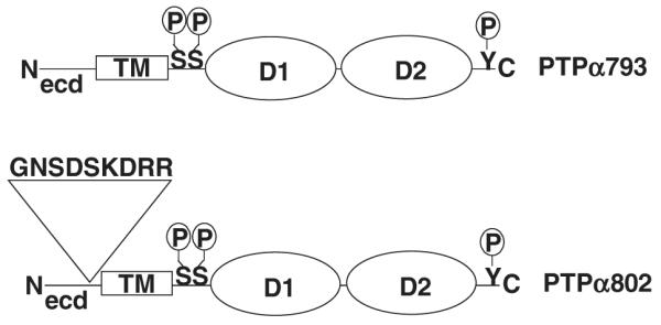
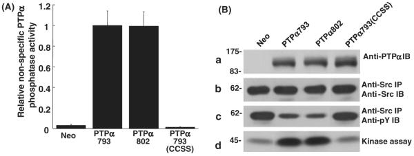
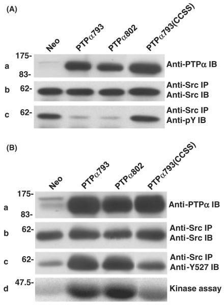
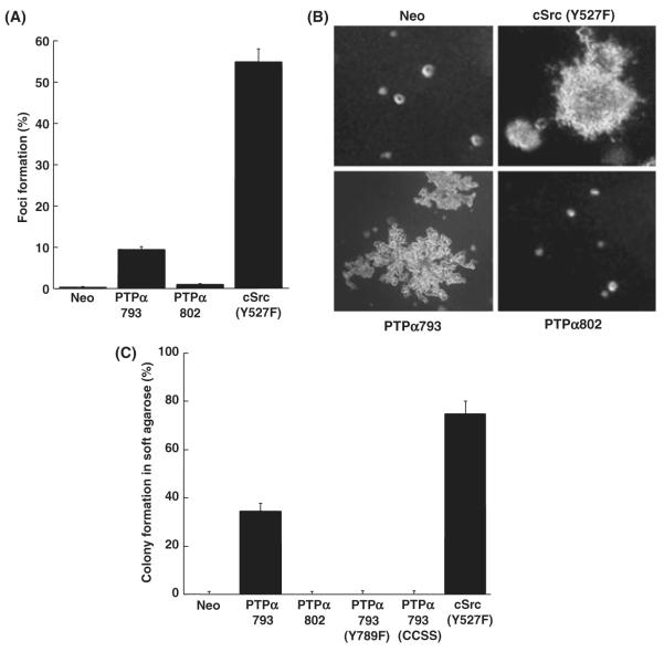
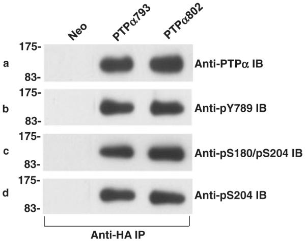
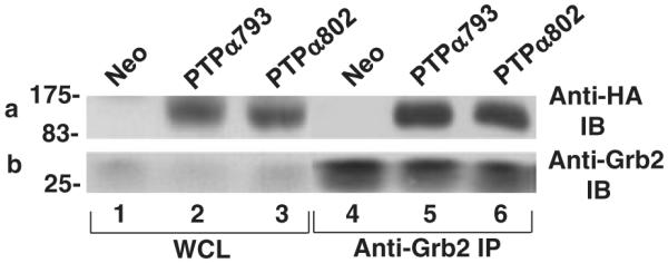
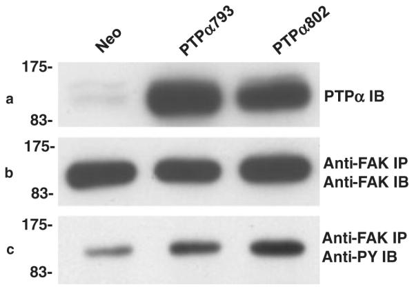
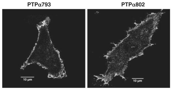
Two isoforms of the transmembrane protein tyrosine phosphatase PTPalpha, which differ by nine amino acids in their extracellular regions, are expressed in a tissue-specific manner. Over-expression of the shorter isoform transforms rodent cells, and it has previously been reasonable to assume that this was a direct consequence of its dephosphorylation and activation of Src. Transformation by the longer wild-type isoform has not previously been studied. We tested the activities of both isoforms in NIH3T3 cells and found that, while both dephosphorylated and activated Src similarly, only the shorter isoform induced focus formation or anchorage-independent growth. Differences in phosphorylation of PTPalpha at its known regulatory sites, Grb2 binding to PTPalpha, phosphorylation level of focal adhesion kinase by PTPalpha, or overall localization were excluded as possible explanations for the differences in transforming activities. The results suggest that transformation by PTPalpha involves at least one function other than, or in addition to, its activation of Src and that this depends on PTPalpha's extracellular domain. Previous studies have suggested that PTPalpha might be a useful target in breast and colon cancer therapy, and the results presented here suggest that it may be advantageous to develop isoform-specific therapeutic reagents.


Figures

Figure 1
Figure 1
Schematic representation of the two isoforms of PTPα. The long form, PTPα802, includes an additional nine amino acids encoded by exon 3 located in the extracellular domain. The locations of the phosphorylation sites Ser180, Ser204 and Tyr789 are indicated. (D1 and D2, catalytic domains; ecd, extracellular domain; TM, transmembrane domain.)
Figure 2
Figure 2
In vitro dephosphorylation of nonspecific and Src substrates by PTPα. (A) Anti-HA immunoprecipitates from lysates from PTPα793, PTPα802 or PTPα793(CCSS) overexpressor cells (induced by removal of doxycycline for 20 h) were incubated with [32P]phosphotyrosine-containing MBP and incubated for 5 or 10 min (in separate experiments to verify reaction linearity) at 30 °C. The amount of [32P] phosphate released was determined by scintillation counting. Immunoblots were used to determine the amount of PTPα present in each reaction and specific activities (relative to that of PTPα793) and standard errors of the mean (n = 3) were computed. (B) Wild-type Src was immunoprecipitated from Src overexpressor cells and subjected to in vitro dephosphorylation by control (Neo) or PTPα proteins that had been immunopurified from induced overexpressor cells. [Equality of the amounts of immunopurified PTPα proteins added to the phosphatase reactions was verified by immunoblotting with anti-PTPα polyclonal antibody (panel a).] The reaction products were divided into three portions that were immunoblotted with either anti-Src (panel b) or anti-phosphotyrosine (panel c) monoclonal antibodies, or used in an in vitro Src kinase assay with [γ-32P]ATP and acid-denatured enolase as substrate (panel d). Src tyrosine phosphorylation was reduced 51 ± 10%, 56 ± 12% or 9 ± 6% (n = 3) by over-expression of PTPα793, PTPα802 or PTPα793(CCSS), respectively; Src kinase activity was increased by factors of 3.4 ± 0.8, 3.6 ± 1.2 or 1.1 ± 0.1 (n = 3; errors are SEMs) by PTPα793, PTPα802 or PTPα793(CCSS), respectively. The positions of molecular weight markers (in kDa) are indicated.
Figure 3
Figure 3
Effect of PTPα over-expression on Src in vivo tyrosine phosphorylation and kinase activity. (A) Neo, PTPα793, PTPα802 and PTPα793(CCSS) overexpressor cells were induced (by removal of doxycycline for 20 h), and lysates were immunoblotted with anti-PTPα polyclonal antibody (panel a) or Src was immunoprecipitated from the lysates and aliquots were immunoblotted with either anti-Src monoclonal antibody (panel b) or anti-pTyr mAb (panel c). Over-expression of PTPα793, PTPα802 or PTPα793(CCSS) decreased tyrosine phosphorylation of Src by 76 ± 7% (n = 4), 79 ± 5% (n = 4) or 19 ± 10% (n = 3), respectively. (B) Lysates from the induced cells were immunoblotted with anti-PTPα polyclonal antibody (panel a) or Src was immunoprecipitated from the lysates and portions were immunoblotted with anti-Src monoclonal antibody (panel b), immunoblotted using anti-dephospho-Y527 Src monoclonal antibody, which reacts only with the activated, Tyr527-dephosphorylated form of Src (panel c), or were subjected to in vitro kinase assay in buffer containing [γ-32P]ATP and acid-denatured enolase (panel d). Over-expression of PTPα793, PTPα802 or PTPα793(CCSS) increased the amount of dephospho-Y527 by factors of 3.8 ± 1.0, 3.9 ± 1.1 or 1.6 ± 0.5 (n = 5) and increased Src kinase activity by factors of 4.9 ± 2.4, 5.9 ± 2.9 or 1.7 ± 0.8 (n = 3), respectively. The positions of molecular weight markers (in kDa) are indicated.
Figure 4
Figure 4
Focus formation and colony formation by PTPα overexpressor cells. (A) Neo (negative control), PTPα793 and PTPα802 NIH3T3-derived overexpressor cells and cSrc(Y527F) overexpressor cells (positive control) were separately (500 cells each) mixed with 2 × 10⁵ NIH3T3 cells and cultured in monolayer without doxycycline for 14 days. The percentages of cells forming foci and standard errors of the mean are shown [two independent experiments each, except six for PTPα802]. [The focus-forming activities of PTPα793 and PTPα802 were statistically different (two-sided t-test) at α = 10−4.] (B) The same cell lines [plus PTPα793(Y789F) and PTPα793(CCSS), not shown] were cultured in 0.3% agarose without doxycycline, and colonies were photographed after 16–18 days. (C) Percentages of cells forming colonies in soft agarose with standard errors of the mean (two independent experiments each, except six for PTPα802). (The colony-forming activities of PTPα793 and PTPα802 were statistically different at α = 10−8).
Figure 5
Figure 5
Phosphorylation status of PTPα proteins. Anti-HA immunoprecipitates from lysates from Neo (control) or PTPα793 or PTPα802 overexpressor cells (induced for 20 h) were divided into four aliquots and immunoblotted with anti-PTPα antibody (panel a), anti-phosphoY789 antibody (panel b), anti-phosphoS180/phosphoS204 antibody (panel c) or anti-phosphoS204 antibody (panel d). No signals were detected with control anti-HA immunoprecipitates from Neo control cells. The ratios between phosphorylation of PTPα793 and PTPα802 at pY789, pS180/pS204 and pS204 were 1.1 ± 0.1, 1.0 ± 0.1 and 1.1 ± 0.1, respectively (n = 4; errors are SEMs). The positions of molecular weight markers (in kDa) are indicated.
Figure 6
Figure 6
Co-immunoprecipitation of PTPα and Grb2. (a) Lysates from induced Neo (control), PTPα793 or PTPα802 overexpressor cells were immunoprecipitated with anti-Grb2 antibody, and the immunoprecipitates were immunoblotted with either anti-HA (panel a) or Grb2 (panel b) antibodies. The positions of molecular weight markers (in kDa) are indicated. The average over six experiments of the ratio of PTPα793-Grb2 binding to PTPα802-Grb2 binding, normalized by the total amounts of PTPα in the cells, was 1.1 ± 0.1.
Figure 7
Figure 7
Regulation of focal adhesion kinase (FAK) phosphorylation by PTPα. Lysates from induced Neo (control), PTPα793, PTPα802 or PTPα793(CCSS) overexpressor cells were immunoblotted with anti-PTPα antibody (panel a) or immunoprecipitated with anti-FAK antibody (panels b and c). Aliquots of the immunoprecipitates were immunoblotted with anti-FAK monoclonal antibody (panel b) or anti-pTyr monoclonal antibody (panel c). FAK tyrosine phosphorylation was increased to roughly equal extents by PTPα793 (4.8 ± 2.6) and PTPα802 (4.65 ± 0.8) in two experiments (errors are SEMs). The positions of molecular weight markers (in kDa) are indicated.
Figure 8
Figure 8
Immunofluorescent localization of overexpressed PTPα793 and PTPα802. PTPα793 and PTPα802 NIH3T3-derived overexpressor cells were induced for 20 h and the subcellular localizations of the HA-tagged PTPα proteins were determined by confocal microscopy as described in Experimental procedures. Ten micron reference bars are shown.

Similar articles

Cited by

References

    1. Ardini E, Agresti R, Tagliabue E, Greco M, Aiello P, Yang LT, Menard S, Sap J. Expression of protein tyrosine phosphatase α (RPTPα) in human breast cancer correlates with low tumor grade, and inhibits tumor cell growth in vitro and in vivo. Oncogene. 2000;19:4979–4987. - PubMed
    1. Autero M, Saharinen J, Pessa-Morikawa T, Soula-Rothhut M, Oetken C, Gassmann M, Bergman M, Alitalo K, Burn P, Gahmberg CG, Mustelin T. Tyrosine phosphorylation of CD45 phosphotyrosine phosphatase by p50csk kinase creates a binding site for p56lck tyrosine kinase and activates the phosphatase. Mol. Cell. Biol. 1994;14:1308–1321. - PMC - PubMed
    1. Blanchetot C, Tertoolen LG, den Hertog J. Regulation of receptor protein-tyrosine phosphatase alpha by oxidative stress. EMBO J. 2002;21:493–503. - PMC - PubMed
    1. Brunton VG, Ozanne BW, Paraskeva C, Frame MC. A role for epidermal growth factor receptor, c-Src and focal adhesion kinase in an in vitro model for the progression of colon cancer. Oncogene. 1997;14:283–293. - PubMed
    1. Buist A, Blanchetot C, Tertoolen LG, den Hertog J. Identification of p130cas as an in vivo substrate of receptor protein-tyrosine phosphatase α. J. Biol. Chem. 2000;275:20754–20761. - PubMed

Publication types

MeSH terms

Substances