

Hes1 regulates embryonic stem cell differentiation by suppressing Notch signaling
- PMID: 20545770
- PMCID: PMC2916211
- DOI: 10.1111/j.1365-2443.2010.01413.x
Hes1 regulates embryonic stem cell differentiation by suppressing Notch signaling
Abstract
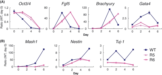
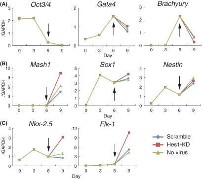
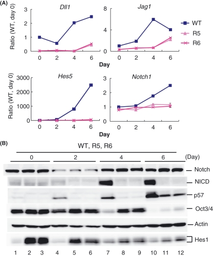
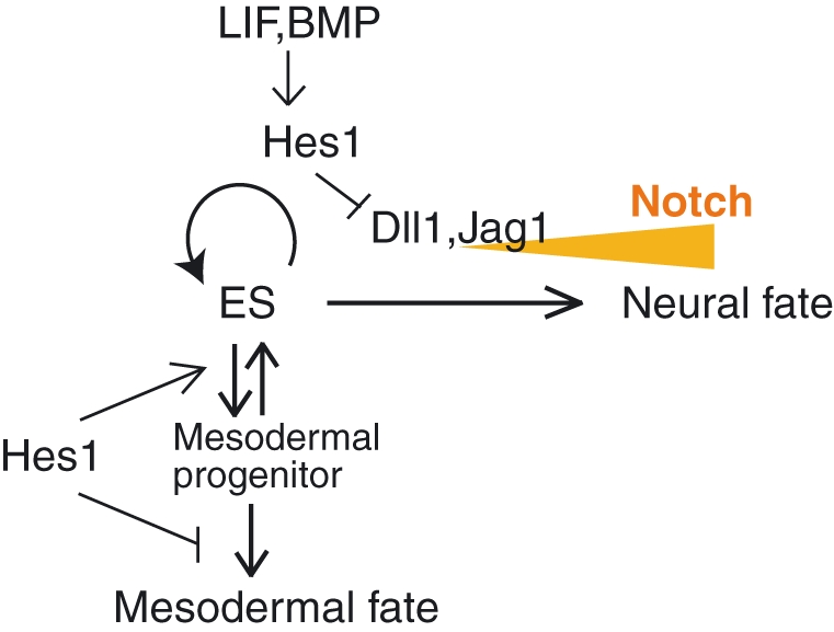
Embryonic stem (ES) cells display heterogeneous responses upon induction of differentiation. Recent analysis has shown that Hes1 expression oscillates with a period of about 3-5 h in mouse ES cells and that this oscillating expression contributes to the heterogeneous responses: Hes1-high ES cells are prone to the mesodermal fate, while Hes1-low ES cells are prone to the neural fate. These outcomes of Hes1-high and Hes1-low ES cells are very similar to those of inactivation and activation of Notch signaling, respectively. These results suggest that Hes1 and Notch signaling lead to opposite outcomes in ES cell differentiation, although they work in the same direction in most other cell types. Here, we found that Hes1 acts as an inhibitor but not as an effector of Notch signaling in ES cell differentiation. Our results indicate that sustained Hes1 expression delays the differentiation of ES cells and promotes the preference for the mesodermal rather than the neural fate by suppression of Notch signaling.
Figures
Similar articles
-
The cyclic gene Hes1 contributes to diverse differentiation responses of embryonic stem cells.Genes Dev. 2009 Aug 15;23(16):1870-5. doi: 10.1101/gad.1823109. Genes Dev. 2009. PMID: 19684110 Free PMC article.
-
Notch-HES1 signaling axis controls hemato-endothelial fate decisions of human embryonic and induced pluripotent stem cells.Blood. 2013 Aug 15;122(7):1162-73. doi: 10.1182/blood-2012-12-471649. Epub 2013 Jun 3. Blood. 2013. PMID: 23733337
-
Suppression of hath1 gene expression directly regulated by hes1 via notch signaling is associated with goblet cell depletion in ulcerative colitis.Inflamm Bowel Dis. 2011 Nov;17(11):2251-60. doi: 10.1002/ibd.21611. Epub 2011 Jan 6. Inflamm Bowel Dis. 2011. PMID: 21987298
-
Expression dynamics and functions of Hes factors in development and diseases.Curr Top Dev Biol. 2014;110:263-83. doi: 10.1016/B978-0-12-405943-6.00007-5. Curr Top Dev Biol. 2014. PMID: 25248479 Review.
-
Rhythmic gene expression in somite formation and neural development.Mol Cells. 2009 May 31;27(5):497-502. doi: 10.1007/s10059-009-0068-1. Epub 2009 May 15. Mol Cells. 2009. PMID: 19466597 Review.
Cited by
-
Peroxisome proliferator-activated receptor γ coactivator-1α (PGC-1α) overexpression alleviates endoplasmic reticulum stress after acute kidney injury.Ren Fail. 2022 Dec;44(1):358-367. doi: 10.1080/0886022X.2022.2035764. Ren Fail. 2022. PMID: 35225153 Free PMC article.
-
Endothelial tip/stalk cell selection requires BMP9-induced βIV-spectrin expression during sprouting angiogenesis.Mol Biol Cell. 2023 Jun 1;34(7):ar72. doi: 10.1091/mbc.E23-02-0064. Epub 2023 Apr 26. Mol Biol Cell. 2023. PMID: 37126382 Free PMC article.
-
Hes1 triggers epithelial-mesenchymal transition (EMT)-like cellular marker alterations and promotes invasion and metastasis of nasopharyngeal carcinoma by activating the PTEN/AKT pathway.Oncotarget. 2015 Nov 3;6(34):36713-30. doi: 10.18632/oncotarget.5457. Oncotarget. 2015. PMID: 26452025 Free PMC article.
-
Activation of CREB-binding protein ameliorates spinal cord injury in tabersonine treatment by suppressing NLRP3/Notch signaling.Arch Med Sci. 2019 Nov 7;19(3):736-743. doi: 10.5114/aoms.2019.89203. eCollection 2023. Arch Med Sci. 2019. PMID: 37313210 Free PMC article.
-
HES factors regulate specific aspects of chondrogenesis and chondrocyte hypertrophy during cartilage development.J Cell Sci. 2016 Jun 1;129(11):2145-55. doi: 10.1242/jcs.181271. Epub 2016 May 9. J Cell Sci. 2016. PMID: 27160681 Free PMC article.
References
-
- Artavanis-Tsakonas S, Rand MD, Lake RJ. Notch signaling: cell fate control and signal integration in development. Science. 1999;284:770–776. - PubMed
-
- Ishibashi M, Ang SL, Shiota K, Nakanishi S, Kageyama R, Guillemot F. Targeted disruption of mammalian hairy and Enhancer of split homolog-1 (HES-1) leads to up-regulation of neural helix-loop-helix factors, premature neurogenesis, and severe neural tube defects. Genes Dev. 1995;9:3136–3148. - PubMed
-
- Ivanova N, Dobrin R, Lu R, Kotenko I, Levorse J, DeCoste C, Schafer X, Lun Y, Lemischka IR. Dissecting self-renewal in stem cells with RNA interference. Nature. 2006;442:533–538. - PubMed
-
- Jang J, Ku SY, Kim JE, et al. Notch inhibition promotes human embryonic stem cell-derived cardiac mesoderm differentiation. Stem Cells. 2008;26:2782–2790. - PubMed
-
- Jensen J, Pedersen EE, Galante P, et al. Control of endodermal endocrine development by Hes-1. Nat. Genet. 2000;24:36–44. - PubMed
Publication types
MeSH terms
Substances
LinkOut - more resources
Full Text Sources
Other Literature Sources
Molecular Biology Databases
