. 2010 Aug;77(3):755-70.
doi: 10.1111/j.1365-2958.2010.07244.x. Epub 2010 Jun 1.

Vibrio cholerae El Tor TcpA crystal structure and mechanism for pilus-mediated microcolony formation

Affiliations

Vibrio cholerae El Tor TcpA crystal structure and mechanism for pilus-mediated microcolony formation

Mindy S Lim et al. Mol Microbiol. 2010 Aug.

Abstract

Type IV pili (T4P) are critical to virulence for Vibrio cholerae and other bacterial pathogens. Among their diverse functions, T4P mediate microcolony formation, which protects the bacteria from host defences and concentrates secreted toxins. The T4P of the two V. cholerae O1 disease biotypes, classical and El Tor, share 81% identity in their TcpA subunits, yet these filaments differ in their interaction patterns as assessed by electron microscopy. To understand the molecular basis for pilus-mediated microcolony formation, we solved a 1.5 A resolution crystal structure of N-terminally truncated El Tor TcpA and compared it with that of classical TcpA. Residues that differ between the two pilins are located on surface-exposed regions of the TcpA subunits. By iteratively changing these non-conserved amino acids in classical TcpA to their respective residues in El Tor TcpA, we identified residues that profoundly affect pilus:pilus interaction patterns and bacterial aggregation. These residues lie on either the protruding d-region of the TcpA subunit or in a cavity between pilin subunits in the pilus filament. Our results support a model whereby pili interact via intercalation of surface protrusions on one filament into depressions between subunits on adjacent filaments as a means to hold V. cholerae cells together in microcolonies.


Figures

Fig. 1
Fig. 1. Comparison of autoagglutination, pilin expression, pilus assembly and TCP:TCP interaction patterns for classical and El Tor V. cholerae biotypes
(A) Autoagglutination of overnight V. cholerae cultures grown under TCP-inducing conditions: wild type classical strain O395 and O395 expressing El Tor TcpA (RT4340) autoagglutinate well (+++) whereas El Tor strain C6706 and the classical mutant strain ML10 (C120A) do not autoagglutinate (-). (B) Immunoblots of TcpA in whole cell cultures (upper panel) and sheared cell supernatant (lower panel) detected with polyclonal anti-TcpA antibody (TcpA6, residues 174-199, (Sun et al., 1991)) to assess TcpA expression and TCP assembly, respectively. (C) Morphology and bundling characteristics of V. cholerae TCP as assessed by negative stain TEM of whole cell cultures (upper panels) and partially purified pili (lower panels). Scale bars, 100 nm.
Fig. 2
Fig. 2. Comparison of the amino acid sequences and atomic structures of classical and El Tor TcpA
(A) Amino acid sequence alignment for mature TcpA proteins from classical V. cholerae O395 (NCBI accession no. ABQ19609) and El Tor strain C6706 (NCBI accession no. AAA85786). The sequences are 81% identical. The secondary structure for TcpACl is indicated above the sequence. The αβ-loop is boxed in green and the D-region is boxed in magenta. α1N (residues 1-28, shaded blue) were replaced by a hexahistidine tag plus a linker region for both pilin crystal structures. α1 is predicted to be a continuous α-helix based on amino acid sequence homology with N. gonorrhoeae and P. aeruginosa pilins, whose full-length structures have been determined (Craig et al., 2003, Craig et al., 2006, Parge et al., 1995). Conserved amino acid differences between classical and El Tor TcpA are shaded in grey and non-conserved differences are shown in black with white letters. Classical-to-El Tor substitutions in the D-region patch are indicated by an asterisk (*) above the corresponding amino acid and the D113A cavity change is indicated by a carat (ˆ). (B)ΔN-TcpAET crystal structure at 1.5 Å resolution, shown as a ribbon diagram. Non-conserved residues between classical and El Tor TcpA are show as stick representations. (C) Superposition of ΔN-TcpAET structure (red) onto ΔN-TcpACl (grey) (Craig et al., 2003). The conserved cysteines are shown in cyan/yellow using ball-and-stick representation.
Fig. 3
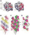
Fig. 3. TcpA electrostatic surface and El Tor TCP filament model
(A) Comparison of the electrostatic surfaces of ΔN-TcpACl and ΔN-TcpAET. The orientation is similar to that shown in Fig. 2C. The electrostatic surface differs in the D-region patch (circled) where there are six non-conserved amino acid differences between the two proteins, each resulting in a change (gain or loss) of a single charge. A second difference is in the location of an aspartate→glycine change at residue 113. (B) Side view of the El Tor TCP filament model generated by superimposing ΔN-TcpAET onto TcpACl in the classical TCP model (Li et al., 2008). The filament is colored to show the subunits arranged in a left-handed three-start helix. (C) Space-filling representation of El Tor TCP to show the positions of the non-conserved amino acids (yellow) in the cavities of the filament and (D) on the protruding D-region. The αβ-loop is colored green and the D-region is magenta in (C) and (D). D-region residues 138, 156, 158, 172, 175 and 187 were selected for classical-to-El Tor mutations, as was cavity residue 113.
Fig. 4
Fig. 4. Analysis of classical-to-El Tor mutants for pilin and pilus expression and pilus morphologies and interaction patterns
(A) Immunoblots of total TcpA in whole cell cultures (upper panel) and TCP in sheared cell supernatant (lower panel) for tcpA mutants and control strains, detected with polyclonal anti-TcpA antibody (TcpA6). All mutants, with the exception of strain ML10, produced TcpA and TCP at or above wild type levels, indicating that the mutations do not aberrantly affect pilin fold or pilus assembly. (B) TEM images of TCP from whole cell cultures of V. cholerae strains harboring the single A156D change, ML15, the complete classical-to-El Tor D-region mutant LC16, the D156A reversion of LC16, strain ML25, and the D113A mutant, ML9. TCP from strains ML15 and ML9 display a mixed pattern of crosshatches and rope-like bundles, whereas LC16 pili are predominantly crosshatched and ML25 TCP form mostly rope-like bundles. Scale bars, 100 nm.
Fig. 5
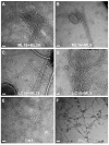
Fig. 5. TEM images of TCP from overnight mixed cultures and from the combined D-region/cavity mutants
Negatively stained pili are from whole cell cultures of (A) ML16 co-inoculated with ML28, (B) ML16 co-inoculated with ML9, (C) LC16 co-inoculated with ML28, (D) LC16 co-inoculated with ML9, and from single inocula combined D-region/cavity mutants (E) DN1 and (F) DN2. Scale bars, 100 nm.

Similar articles

Cited by

References

    1. Anantha RP, Stone KD, Donnenberg MS. Role of BfpF, a member of the PilT family of putative nucleotide-binding proteins, in type IV pilus biogenesis and in interactions between enteropathogenic Escherichia coli and host cells. Infect Immun. 1998;66:122–131. - PMC - PubMed
    1. Barken KB, Pamp SJ, Yang L, Gjermansen M, Bertrand JJ, Klausen M, et al. Roles of type IV pili, flagellum-mediated motility and extracellular DNA in the formation of mature multicellular structures in Pseudomonas aeruginosa biofilms. Environ Microbiol. 2008;10:2331–2343. - PubMed
    1. Biais N, Ladoux B, Higashi D, So M, Sheetz M. Cooperative retraction of bundled type IV pili enables nanonewton force generation. PLoS Biol. 2008;6:e87. - PMC - PubMed
    1. Bieber D, Ramer SW, Wu CY, Murray WJ, Tobe T, Fernandez R, Schoolnik GK. Type IV pili, transient bacterial aggregates, and virulence of enteropathogenic Escherichia coli. Science. 1998;280:2114–2118. - PubMed
    1. Brunger AT, Adams PD, Clore GM, DeLano WL, Gros P, Grosse-Kunstleve RW, et al. Crystallography and NMR system: A new software suite for macromolecular structure determination. Acta Crystallogr D Biol Crystallogr. 1998;54(Pt 5):905–921. - PubMed

Publication types

MeSH terms

Substances

Associated data