. 2010 Aug;77(3):658-71.
doi: 10.1111/j.1365-2958.2010.07236.x. Epub 2010 Jun 1.

Trypanosoma brucei pteridine reductase 1 is essential for survival in vitro and for virulence in mice

Affiliations
Free PMC article

Trypanosoma brucei pteridine reductase 1 is essential for survival in vitro and for virulence in mice

Natasha Sienkiewicz et al. Mol Microbiol. 2010 Aug.
Free PMC article

Abstract

Gene knockout and knockdown methods were used to examine essentiality of pteridine reductase (PTR1) in pterin metabolism in the African trypanosome. Attempts to generate PTR1 null mutants in bloodstream form Trypanosoma brucei proved unsuccessful; despite integration of drug selectable markers at the target locus, the gene for PTR1 was either retained at the same locus or elsewhere in the genome. However, RNA interference (RNAi) resulted in complete knockdown of endogenous protein after 48 h, followed by cell death after 4 days. This lethal phenotype was reversed by expression of enzymatically active Leishmania major PTR1 in RNAi lines ((oe)RNAi) or by addition of tetrahydrobiopterin to cultures. Loss of PTR1 was associated with gross morphological changes due to a defect in cytokinesis, resulting in cells with multiple nuclei and kinetoplasts, as well as multiple detached flagella. Electron microscopy also revealed increased numbers of glycosomes, while immunofluorescence microscopy showed increased and more diffuse staining for glycosomal matrix enzymes, indicative of mis-localisation to the cytosol. Mis-localisation was confirmed by digitonin fractionation experiments. RNAi cell lines were markedly less virulent than wild-type parasites in mice and virulence was restored in the (oe)RNAi line. Thus, PTR1 may be a drug target for human African trypanosomiasis.


Figures

Fig. 1
Fig. 1
The effect of RNAi-dependent depletion of PTR1 on growth in the presence or absence of tetrahydrobiopterin. A. Effect of RNAi on cell growth. Four independent RNAi clones were analysed for growth defects following induction with tetracycline in the absence of tetrahydrobiopterin. WT (open circles) and RNAi cells (non-induced cells – open squares and induced cells – closed squares) were seeded at 1 × 10⁴ cells ml−1 and growth was determined over a 2 week period. WT, non-induced and induced cells (1 × 10⁶ cells per lane) were harvested at 0 (non-induced), 24 h and 48 h (induced) and processed for Western blot analysis. The blot was sequentially probed with anti-TbPTR1 (inset, lower panel) and anti-TbBIP (inset, upper panel) as loading control. B. Effect of tetrahydrobiopterin on cell growth. WT cells and a freshly derived RNAi clone were seeded into FDM or HMI9-T supplemented with or without 10 µM tetrahydrobiopterin at 1 × 10⁴ cells ml−1 and growth monitored over 1 week. Symbols: WT (open circle; no additions), non-induced RNAi cells (closed circles) and tetracycline-induced cells plus or minus tetrahydrobiopterin (closed and open squares respectively). Data shown for FDM only; HMI9-T shows essentially the same effect.
Fig. 3
Fig. 3
PTR1 enzyme activity in WT and transgenic T. brucei. Cell lysates were assayed for PTR1 activity at various protein concentrations as described in the materials and methods. WT, open circles; SKO, closed circles; oeRNAi minus tetracycline, open squares; oeRNAi plus tetracycline, closed squares.
Fig. 2
Fig. 2
Rescue of RNAi growth defect by expression of LmPTR1. A. Cumulative growth of WT (open circles) non-induced oeRNAi cells (open inverted triangles) and tetracycline-induced oeRNAi cells (closed triangles). B. Western blot analysis demonstrating knockdown of TbPTR1 in oeRNAi cells. Non-induced (-) and 48 h tetracycline induced (+) cells were probed sequentially with anti-TbPTR1 and anti-LmPTR1. A 55 kDa non-specific band was detected with anti-TbPTR1, which was not effect by the knockdown of PTR1 and serves as an internal loading control (top panel), while LmPTR1 is present in both (bottom panel).
Fig. 4
Fig. 4
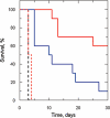
Virulence phenotype of PTR1 mutants in mice. Groups of five mice were infected with WT, non-induced cells for RNAi and oeRNAi cells and parasitaemia monitored at intervals over a 30 day period. The Kaplan-Meier survival graph shows the aggregated results of two independent experiments. Symbols: WT infection, no doxycycline (black dashed line); RNAi infection, no doxycycline (blue solid line); RNAi infection plus doxycycline (red solid line); oeRNAi infection, no doxycycline (red dashed line); oeRNAi infection with doxycycline (black dashed line, identical to WT infection).
Fig. 5
Fig. 5
PTR1 depletion results in changes to cell cycle progression. Microscopic analysis of DAPI stained RNAi cells of non-induced (0 h) and induced (24, 48 and 72 h) populations analysed to determine the proportion of trypanosomes with different numbers of kinetoplasts (K) and nuclei (N). Percentage bar chart depicts changes in the K/N content per cell after tetracycline induction of RNAi. Approximately 300 parasites per population were analysed (n = 2, ± SD): non-induced control (black bars); 24 h induction (dark grey bars); 48 h induction (light grey bars) and 72 h induction (white bars).
Fig. 6
Fig. 6
Morphology of PTR1-depleted cells by electron microscopy. A. TEM section of non-induced controls. B and C. TEM sections of 72-h induced cells depicting increased number of flagella and glycosomes. D. SEM images of 72-h induced cells, one with a normal (top left) and abnormal (bottom right) appearance compared with non-induced control (inset). E. Equivalent DAPI-stained 72-h induced cell with multiple flagella. Abbreviations used in TEM sections: nucleus (N); nuclear membrane (NM); basal bodies (BB); kinetoplast (K); flagellar rod (FR); flagellar pocket (FP); axonemes (Ax and black arrow heads); glycosomes (G); lysosomes (L); paraxial rod (PR); microtubules (MT); acidocalcisomes (Ac); autophagic vacuoles (AV) internal non-membrane bound flagellum (*). White arrow heads in SEM images highlight flagella. Black and white bars represent 200 nm.
Fig. 7
Fig. 7
Localization and distribution of GAPDH by immuno-gold labelling. Thin-layer sections were labelled with anti-GAPDH and stained with protein A gold particles and examined by TEM. WT (A), RNAi non-induced (B) and induced (C–D) at 72 h. Abbreviations used: nucleus (N); glycosome (G) and white asterisks alongside black dots represent 10 nm gold particles present within glycosomes, while white arrows depict particles outside glycosomes. Scale bar represents 200 nm.
Fig. 9
Fig. 9
Immunolocalisation of aldolase and paraflagellar rod marker, ROD1 in RNAi depleted cells. Co-localization of aldolase (glycosomal marker, green) and ROD1 (paraflagellar rod marker, red) with DAPI (blue) on RNAi cells. Non-induced (A); induced 48 h (B); and 72 h (C) RNAi cells. MN, multinucleated, MK, multikinetoplastid, and scale bar represents 10 µm.
Fig. 8
Fig. 8
The effect of PTR1 depletion on the subcellular distribution of the glycosomal matrix enzyme, GAPDH by immunofluorescence microscopy. Non-induced (A), induced 48 h (B) and 72 h (C) RNAi cells were labelled with a glycosomal matrix marker, GAPDH (green) with staining of nuclear and kinetoplast DNA by DAPI (blue). MN, multinucleated, MK, multikinetoplastid. Scale bar represents 10 µm.
Fig. 10
Fig. 10
The effect of PTR1 depletion on the subcellular distribution of glycolytic enzymes. Digitonin fractionation of intact WT and RNAi cells grown for 48 h (in the absence ‘–’ and presence ‘+’ of tetracycline) were incubated with 150 µg ml−1 of digitonin and processed as described in Material and Methods. The release of PTR1, enolase, PEX13 and GAPDH from digitonin-treated cells was determined in supernatant (S) and pellet (P) fractions by Western blot.

Similar articles

Cited by

References

    1. Alibu VP, Storm L, Haile S, Clayton C, Horn D. A doubly inducible system for RNA interference and rapid RNAi plasmid construction in Trypanosoma brucei. Mol Biochem Parasitol. 2005;139:75–82. - PubMed
    1. Armarego WLF, Randles D, Taguchi H. Km and kcat values for [6,6,7,7-2H]7,8(6H)-dihydropterin and 2,6-diamino-5-iminopyrimidin-4-one with dihydropteridine reductase. Biochem J. 1983;211:357–361. - PMC - PubMed
    1. Banerjee SK, Kessler PS, Saveria T, Parsons M. Identification of trypanosomatid PEX19: functional characterization reveals impact on cell growth and glycosome size and number. Mol Biochem Parasitol. 2005;142:47–55. - PubMed
    1. Bello AR, Nare B, Freedman D, Hardy L, Beverley SM. PTR1: a reductase mediating salvage of oxidized pteridines and methotrexate resistance in the protozoan parasite Leishmania major. Proc Natl Acad Sci USA. 1994;91:11442–11446. - PMC - PubMed
    1. Berriman M, Ghedin E, Hertz-Fowler C, Blandin G, Renauld H, Bartholomeu DC, et al. The Genome of the African Trypanosome Trypanosoma brucei. Science. 2005;309:416–422. - PubMed

Publication types

LinkOut - more resources