M. tuberculosis intramembrane protease Rip1 controls transcription through three anti-sigma factor substrates
- PMID: 20545848
- PMCID: PMC3008510
- DOI: 10.1111/j.1365-2958.2010.07232.x
M. tuberculosis intramembrane protease Rip1 controls transcription through three anti-sigma factor substrates
Abstract
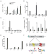
Regulated intramembrane proteolysis (RIP) is a mechanism of transmembrane signal transduction that functions through intramembrane proteolysis of substrates. We previously reported that the RIP metalloprotease Rv2869c (Rip1) is a determinant of Mycobacterium tuberculosis (Mtb) cell envelope composition and virulence, but the substrates of Rip1 were undefined. Here we show that Rip1 cleaves three transmembrane anti-sigma factors: anti-SigK, anti-SigL and anti-SigM, negative regulators of Sigma K, L and M. We show that transcriptional activation of katG in response to phenanthroline requires activation of SigK and SigL by Rip1 cleavage of anti-SigK and anti-SigL. We also demonstrate a Rip1-dependent pathway that activates the genes for the mycolic acid biosynthetic enzyme KasA and the resuscitation promoting factor RpfC, but represses the bacterioferritin encoding gene bfrB. Regulation of these three genes by Rip1 is not reproduced by deletion of Sigma K, L or M, either indicating a requirement for multiple Rip1 substrates or additional arms of the Rip1 pathway. These results identify a branched proteolytic signal transduction system in which a single intramembrane protease cleaves three anti-sigma factor substrates to control multiple downstream pathways involved in lipid biosynthesis and defence against oxidative stress.
Figures


Similar articles
-
The Rip1 protease of Mycobacterium tuberculosis controls the SigD regulon.J Bacteriol. 2014 Jul;196(14):2638-45. doi: 10.1128/JB.01537-14. Epub 2014 May 9. J Bacteriol. 2014. PMID: 24816608 Free PMC article.
-
Site-2 protease substrate specificity and coupling in trans by a PDZ-substrate adapter protein.Proc Natl Acad Sci U S A. 2013 Nov 26;110(48):19543-8. doi: 10.1073/pnas.1305934110. Epub 2013 Nov 11. Proc Natl Acad Sci U S A. 2013. PMID: 24218594 Free PMC article.
-
The Rip1 intramembrane protease contributes to iron and zinc homeostasis in Mycobacterium tuberculosis.mSphere. 2023 Aug 24;8(4):e0038922. doi: 10.1128/msphere.00389-22. Epub 2023 Jun 15. mSphere. 2023. PMID: 37318217 Free PMC article.
-
Regulated proteolysis: control of the Escherichia coli σ(E)-dependent cell envelope stress response.Subcell Biochem. 2013;66:129-60. doi: 10.1007/978-94-007-5940-4_6. Subcell Biochem. 2013. PMID: 23479440 Review.
-
Regulated intramembrane proteolysis in the control of extracytoplasmic function sigma factors.Res Microbiol. 2009 Nov;160(9):696-703. doi: 10.1016/j.resmic.2009.08.019. Epub 2009 Sep 22. Res Microbiol. 2009. PMID: 19778605 Review.
Cited by
-
Genome-wide screen identifies host loci that modulate Mycobacterium tuberculosis fitness in immunodivergent mice.G3 (Bethesda). 2023 Aug 30;13(9):jkad147. doi: 10.1093/g3journal/jkad147. G3 (Bethesda). 2023. PMID: 37405387 Free PMC article.
-
Substrate specificity of MarP, a periplasmic protease required for resistance to acid and oxidative stress in Mycobacterium tuberculosis.J Biol Chem. 2013 May 3;288(18):12489-99. doi: 10.1074/jbc.M113.456541. Epub 2013 Mar 15. J Biol Chem. 2013. PMID: 23504313 Free PMC article.
-
Genome-wide screen identifies host loci that modulate M. tuberculosis fitness in immunodivergent mice.bioRxiv [Preprint]. 2023 Mar 6:2023.03.05.528534. doi: 10.1101/2023.03.05.528534. bioRxiv. 2023. Update in: G3 (Bethesda). 2023 Aug 30;13(9):jkad147. doi: 10.1093/g3journal/jkad147. PMID: 36945430 Free PMC article. Updated. Preprint.
-
Mycobacterium tuberculosis Hip1 modulates macrophage responses through proteolysis of GroEL2.PLoS Pathog. 2014 May 15;10(5):e1004132. doi: 10.1371/journal.ppat.1004132. eCollection 2014 May. PLoS Pathog. 2014. PMID: 24830429 Free PMC article.
-
The role of site-2-proteases in bacteria: a review on physiology, virulence, and therapeutic potential.Microlife. 2023 May 3;4:uqad025. doi: 10.1093/femsml/uqad025. eCollection 2023. Microlife. 2023. PMID: 37223736 Free PMC article. Review.
References
Publication types
MeSH terms
Substances
Grants and funding
LinkOut - more resources
Full Text Sources
Miscellaneous

