. 2010 Aug;77(3):730-42.
doi: 10.1111/j.1365-2958.2010.07243.x. Epub 2010 Jun 10.

Mycobacterium tuberculosis CYP125A1, a steroid C27 monooxygenase that detoxifies intracellularly generated cholest-4-en-3-one

Affiliations

Mycobacterium tuberculosis CYP125A1, a steroid C27 monooxygenase that detoxifies intracellularly generated cholest-4-en-3-one

Hugues Ouellet et al. Mol Microbiol. 2010 Aug.

Abstract

The infectivity and persistence of Mycobacterium tuberculosis requires the utilization of host cell cholesterol. We have examined the specific role of cytochrome P450 CYP125A1 in the cholesterol degradation pathway using genetic, biochemical and high-resolution mass spectrometric approaches. The analysis of lipid profiles from cells grown on cholesterol revealed that CYP125A1 is required to incorporate the cholesterol side-chain carbon atoms into cellular lipids, as evidenced by an increase in the mass of the methyl-branched phthiocerol dimycocerosates. We observed that cholesterol-exposed cells lacking CYP125A1 accumulate cholest-4-en-3-one, suggesting that this is a physiological substrate for this enzyme. Reconstitution of enzymatic activity with spinach ferredoxin and ferredoxin reductase revealed that recombinant CYP125A1 indeed binds both cholest-4-en-3-one and cholesterol, efficiently hydroxylates both of them at C-27, and then further oxidizes 27-hydroxycholest-4-en-3-one to cholest-4-en-3-one-27-oic acid. We determined the X-ray structure of cholest-4-en-3-one-bound CYP125A1 at a resolution of 1.58 A. CYP125A1 is essential for growth of CDC1551 in media containing cholesterol or cholest-4-en-3-one. In its absence, the latter compound is toxic for both CDC1551 and H37Rv when added with glycerol as a second carbon source. CYP125A1 is a key enzyme in cholesterol metabolism and plays a crucial role in circumventing the deleterious effect of cholest-4-en-3-one.


Figures
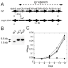
Fig. 1
Fig. 1
Inactivation of cyp125A1 and growth characteristics. (A) Schematic representation of the igr operon consisting of cyp125A1 and five other genes before and after insertion of the hyg resistance gene in cyp125A1. (B) Southern blot of EcoR I-digested genomic DNA showing the hybridizing fragment of WT (1.5 kb) and of two cyp125A1 deletion mutants, HO1 and HO2 (5.5 kb). (C) Representative growth curves are shown of WT (circle), Δcyp125A1 (HO1, square), and Δcyp125A1/cyp125A1 (HO11, diamond) strains on minimal media containing glycerol (open symbols) or cholesterol (filled symbols). The growth was monitored by measuring the absorbance at 600 nm as a function of time.
Fig. 2
Fig. 2
Mass spectrometric analysis of PDIM extracted from the WT, HO1 and HO11 strains. Cells were incubated with glycerol, cholesterol or heptadeuterated-cholesterol for 24 h in 7H9 medium supplemented with 0.5% bovine serum albumin, 0.085% NaCl and 0.05% Tween-80. Apolar lipids were prepared and monitored by LTQ-FT/MS as described in the Experimental Procedures section. Addition of cholesterol leads to an increase in the mass of the PDIM.
Fig. 3
Fig. 3
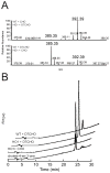
Accumulation of a cholesterol-derived metabolite in M. tuberculosis cells. (A) Co-injection (1:1) of apolar lipid extracts prepared from WT and HO1 cells incubated with cholesterol and heptadeuterated-cholesterol, respectively (top panel), and vice-versa (bottom panel). The m/z value for the non-deuterated metabolite is 385.35, while that of the corresponding heptadeuterated metabolite is 392.39. (B) HPLC traces showing absorbance at 240 nm versus time for separation of crude apolar lipids extracted from HO1 cells grown on cholesterol and of authentic cholest-4-en-3-one as a reference. The retention time for the major peak was ~24.2 min. The traces for separation of crude apolar lipids extracted from WT and HO1 cells grown on heptadeuterated cholesterol are offset for clarity.
Fig. 4
Fig. 4
Binding of cholest-4-en-3-one and cholesterol to recombinant CYP125A1. (A) Absolute Soret region absorption spectrum of CYP125A1 protein (12.95 μM heme) in its resting (thin line), cholest-4-en-3-one (dashed line) cholesterol-bound (thick line) forms. The concentration dependence of ligand binding was deduced from the difference absorption changes obtained from the titration of protein (10 μM P450) with increasing concentrations of cholest-4-en-3-one (B) and cholesterol (C). Representative sets of the difference spectra that were recorded are shown in the insets. Spectrophotometric titrations were carried out as described in the Experimental Procedures section.
Fig. 5
Fig. 5
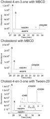
Oxidation of cholest-4-en-3-one and cholesterol by CYP125A1. Offset GC traces for incubation of cholest-4-en-3-one (top and bottom panels) or cholesterol (middle panel) with CYP125A1 in reconstituted enzymatic activity assays. For the top and middle panels, the reactions were performed in the presence of 0.45% (w/v) MBCD, while that of the bottom panel was performed in the presence of 0.05% (v/v) Tween-20. The peak labeled S corresponds to the substrate cholest-4-en-3-one or cholesterol. The peaks labeled -CH2OH, –CHO and -COOH correspond to the products 27-hydroxy cholest-4-en-3-one, 27-al cholest-4-en-3-one TMS ethers, and 27-carboxylic acid cholest-4-en-3-one TMS ester (after derivatization for GC-MS), respectively. The inset of the bottom panel is a ~5x enlargement of product peaks region.
Fig. 6
Fig. 6
Substrate binding tunnel. View of the cholest-4-en-3-one-bound CYP125A1 clipped by a plane through the substrate binding tunnel orthogonal (A) or parallel (B) to the plane of the tetracyclic sterol nucleus. Hydrophobic areas are orange, hydrophilic areas are blue. The residues preventing cholest-4-en-3-one from sliding toward the heme are labelled. The cholest-4-en-3-one color scheme is carbon grey, oxygen red. The image was generated using CHIMERA (Pettersen et al., 2004).
Fig. 7
Fig. 7
Cholest-4-en-3-one binding. Stereoscopic view of cholest-4-en-3-one (highlighted in yellow) surrounded by the CYP125A1 side chains (cyan) within 4 Å. Main chain atoms are shown for a couple of consecutive residues P213-K214 constituting a crevice where the sterol A-ring resides. 2Fo-Fc electron density map (grey mesh) was generated with the cholest-4-en-3-one coordinates omitted. For clarity, heme, Q112 and V115 are excluded from map calculation. Oxygen is coloured red, nitrogen blue, heme orange. The image was generated using PYMOL (DeLano, 2002).
Fig. 8
Fig. 8
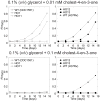
Growth inhibition by cholest-4-en-3-one. Representative growth curves on minimal media with 0.1% (v/v) glycerol in combination with 0.01 mM cholest-4-en-3-one (top panels) or 0.1 mM cholest-4-en-3-one (bottom panels). The growth was monitored by measuring the absorbance at 600 nm in function of time.

Similar articles

Cited by

References

    1. (WHO), W. H. O. World Health Organization Facts Sheets on Tuberculosis 2009
    1. Appleby CA. Purification of Rhizobium cytochromes P-450. Methods Enzymol. 1978;52:157–166. - PubMed
    1. Bardarov S, Bardarov S, Jr, Pavelka MS, Jr, Sambandamurthy V, Larsen M, Tufariello J, Chan J, Hatfull G, Jacobs WR., Jr Specialized transduction: an efficient method for generating marked and unmarked targeted gene disruptions in Mycobacterium tuberculosis, M. bovis BCG and M. smegmatis. Microbiology. 2002;148:3007–3017. - PubMed
    1. Boshoff HI, Barry CE., 3rd A low-carb diet for a high-octane pathogen. Nat Med. 2005a;11:599–600. - PubMed
    1. Boshoff HI, Barry CE., 3rd Tuberculosis - metabolism and respiration in the absence of growth. Nat Rev Microbiol. 2005b;3:70–80. - PubMed

Publication types

MeSH terms