. 2010 Aug;63(4):636-50.
doi: 10.1111/j.1365-313X.2010.04268.x.

Identification of protein stability determinants in chloroplasts

Affiliations
Free PMC article

Identification of protein stability determinants in chloroplasts

Wiebke Apel et al. Plant J. 2010 Aug.
Free PMC article

Abstract

Although chloroplast protein stability has long been recognised as a major level of post-translational regulation in photosynthesis and gene expression, the factors determining protein stability in plastids are largely unknown. Here, we have identified stability determinants in vivo by producing plants with transgenic chloroplasts that express a reporter protein whose N- and C-termini were systematically modified. We found that major stability determinants are located in the N-terminus. Moreover, testing of all 20 amino acids in the position after the initiator methionine revealed strong differences in protein stability and indicated an important role of the penultimate N-terminal amino acid residue in determining the protein half life. We propose that the stability of plastid proteins is largely determined by three factors: (i) the action of methionine aminopeptidase (the enzyme that removes the initiator methionine and exposes the penultimate N-terminal amino acid residue), (ii) an N-end rule-like protein degradation pathway, and (iii) additional sequence determinants in the N-terminal region.


Figures

Figure 1
Figure 1
Construction of transformation vectors to analyse determinants of protein stability in plastids.(a) Physical map of the targeting region in the plastid genome from which the chloroplast transformation vectors were derived. The transgenes are targeted to the intergenic spacer between the trnfM and trnfG genes. Restriction sites used for RFLP analysis and their corresponding fragment sizes in the wild-type are indicated. Hybridisation probes are indicated by horizontal bars. Genes above the line are transcribed from the left to the right, those below the line are transcribed in the opposite direction.(b, c) Maps of the vector used to test N-terminal (b) and C-terminal (c) determinants of protein stability. Ins-N represents the sequence for the eight or nine N-terminal amino acids of five chloroplast proteins. Ins-C represents the sequence for the eight C-terminal amino acids of five chloroplast proteins. Relevant restriction sites used for cloning and/or exchange of N- or C-terminal sequence motifs are indicated. Vectors pWA-N and pWA-C harbour the selectable marker gene aadA (Svab and Maliga, 1993) driven by a chimeric rRNA operon promoter (Prrn) and the 3′ UTR from the psbA gene (TpsbA). The gfp reporter gene is controlled by the ribosomal RNA operon promoter (Prrn), a 5′ UTR containing an rbcL-derived Shine–Dalgarno sequence, and the 3′ UTR from the rps16 gene (T3′rps16).(d) Schematic representation of the N- and C-terminal fusion constructs, with the amino acid sequences fused to GFP given using the three-letter code. The codon in the PsbE N-terminus that was systematically modified is boxed. For technical reasons (codon constraints), the PGK N-terminus comprises nine amino acids.
Figure 2
Figure 2
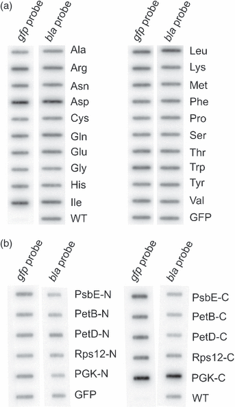
Analysis of RNA accumulation in E. coli by slot-blot assays. (a) Constructs harbouring mutations of the penultimate N-terminal amino acid residue.(b) Constructs for testing N- and C-terminal determinants of protein stability. Samples of 100 ng total RNA were blotted and hybridised to a gfp-specific probe, and, as a control for equal loading, to a bla-specific probe detecting transcripts from the ampicillin resistance gene localised on the same plasmid vector.WT, negative control (a bacterial strain transformed with a control vector lacking a gfp gene); GFP, positive control (a bacterial strain expressing unfused GFP).
Figure 3
Figure 3
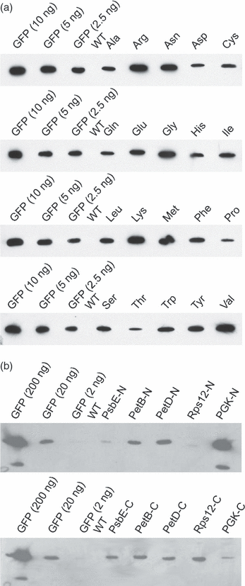
Western blot analysis to determine accumulation of the GFP reporter in E. coli using an anti-GFP antibody.(a) Constructs harbouring mutations of the penultimate N-terminal amino acid residue. For each transformed strain, 6 μg total soluble protein was loaded.(b) Constructs for testing N- and C-terminal determinants of protein stability. For each construct, 8 μg total soluble protein was loaded.WT, protein extract from bacteria transformed with a control vector lacking a gfp gene. For quantitative assessment of protein accumulation, a dilution series of purified recombinant GFP was included. For clarity, all GFP-containing samples are separated from each other by an empty lane. Data were confirmed by analysis of three biological replicates. Equal loading was further confirmed by Coomassie staining of the high-molecular-weight region of the gel (which was not blotted). To control for technical variation, the experiments were repeated three times and similar results were obtained.
Figure 4
Figure 4
Molecular and genetic analysis of transplastomic tobacco lines.(a) Southern blot analysis of transplastomic lines generated with constructs harbouring mutations of the penultimate N-terminal amino acid residue. Total cellular DNA was digested with BglII and hybridised to a radiolabelled probe detecting the region of the plastid genome that flanks the transgene insertion site (see Figure 1a). WT, wild-type control. Fragment sizes of the molecular weight marker are indicated at the right in kb.(b) Southern blot analysis of transplastomic lines produced to test N- and C-terminal determinants of protein stability. DNA samples were digested with XcmI. The sizes of hybridising bands are indicated on the right (in kb). The size difference of 2.3 kb between the hybridisation signals in the wild-type and the transplastomic lines corresponds to the combined size of the two transgene cassettes (Figure 1a). WT, wild-type control. Fragment sizes of the molecular weight marker are indicated at the right in kb.(c) Representative examples of seed tests to confirm homoplasmy of transplastomic lines. Seeds from the wild-type (WT) and seven selfed transplastomic plants were germinated on medium with spectinomycin. The lack of segregation of the antibiotic resistance in the T1 generation confirms the homoplasmic state of all transplastomic lines.
Figure 5
Figure 5
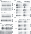
Analysis of transcript accumulation and ribosome association of mRNAs from gfp fusion genes in plastids.(a) RNA accumulation in transplastomic plants transformed with constructs harbouring mutations of the penultimate N-terminal amino acid residue.(b) RNA accumulation in plants transformed with constructs for testing N- and C-terminal determinants of protein stability. To control for equal loading, blots were first hybridised to a gfp probe, then stripped and re- hybridised to a 16S rRNA probe. The gfp probe detects two major transcript species. The lower band (0.9 kb) represents mature monocistronic gfp mRNA, and the upper band most likely represents a stable read-through transcript (Zhou et al., 2007, 2008).(c) Analysis of polysome loading in selected transplastomic lines generated with constructs harbouring mutations of the penultimate N-terminal amino acid residue.(d) Analysis of polysome loading in selected transplastomic lines transformed with constructs for testing N- and C-terminal determinants of protein stability.Collected fractions from the sucrose density gradients were numbered from the top to the bottom. Ribosome distribution in the gradients is revealed by hybridisation to a 16S rRNA probe. Equal aliquots of extracted RNAs from all fractions were separated by denaturing agarose gel electrophoresis, blotted and hybridised to radiolabelled probes specific for gfp and 16S rRNA. The wedges above each blot indicate the gradient in sucrose density (from low to high). As a control, a sample was treated with puromycin to cause dissociation of ribosomes from the mRNAs. To control for technical variation, the experiments were repeated three times and identical results were obtained.
Figure 7
Figure 7
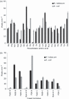
Quantification of GFP accumulation in E. coli and tobacco plastids.Three biological replicates of all blots were quantified using imagej software (http://rsb.info.nih.gov/ij/). The standard deviation is indicated by error bars.(a) Protein accumulation in plants transformed with constructs harbouring mutations of the penultimate N-terminal amino acid residue.(b) Protein accumulation in plants transformed with constructs for testing N- and C-terminal determinants of protein stability.
Figure 6
Figure 6
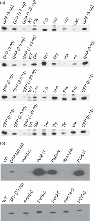
Western blot analysis to determine GFP accumulation in transplastomic tobacco plants using an anti-GFP antibody.(a) GFP accumulation in plants transformed with constructs harbouring mutations of the penultimate N-terminal amino acid residue. For each plant line, 20 μg total soluble protein was loaded. For quantitative assessment of protein accumulation, purified recombinant GFP was loaded at two dilutions.(b) GFP accumulation in plants transformed with constructs for testing N- and C-terminal determinants of protein stability. For each plant line, 5 μg total soluble protein was loaded. For clarity, all GFP-containing samples were separated from each other by an empty lane. The reproducibility of the data was confirmed by analysis of three biological replicates. Equal loading was further confirmed by Coomassie staining of the high-molecular-weight region of the gel (which was not blotted) and assessing the amount of large subunit of Rubisco (RbcL). WT, wild-type tobacco.
Figure 8
Figure 8
Protein stability with respect to the N-termini of plastid proteins.(a) Frequency of occurrence of each of the 20 proteinogenic amino acids after the initiator methionine in plastid-encoded proteins.(b) Relationship between protein stability and the frequency of occurrence of the 20 proteinogenic amino acids as penultimate N-terminal residues in plastid proteins.

References

    1. Adam Z. Protein stability and degradation in chloroplasts. Plant Mol. Biol. 1996;32:773–783. - PubMed
    1. Adam Z. Chloroplast proteases: Possible regulators of gene expression. Biochimie. 2000;82:647–654. - PubMed
    1. Adam Z. Protein stability and degradation in plastids. Top. Curr. Genet. 2007;19:315–338.
    1. Adam Z, Rudella A, van Wijk KJ. Recent advances in the study of Clp, FtsH and other proteases located in chloroplasts. Curr. Opin. Plant Biol. 2006;9:234–240. - PubMed
    1. Barber J, Nield J, Morris EP, Zheleva D, Hankamer B. The structure, function and dynamics of photosystem two. Physiol. Plant. 1997;100:817–827.

Publication types

MeSH terms