. 2010 Sep;11(9):1151-67.
doi: 10.1111/j.1600-0854.2010.01085.x. Epub 2010 Jun 2.

Zebrafish class 1 phosphatidylinositol transfer proteins: PITPbeta and double cone cell outer segment integrity in retina

Affiliations

Zebrafish class 1 phosphatidylinositol transfer proteins: PITPbeta and double cone cell outer segment integrity in retina

Kristina E Ile et al. Traffic. 2010 Sep.

Abstract

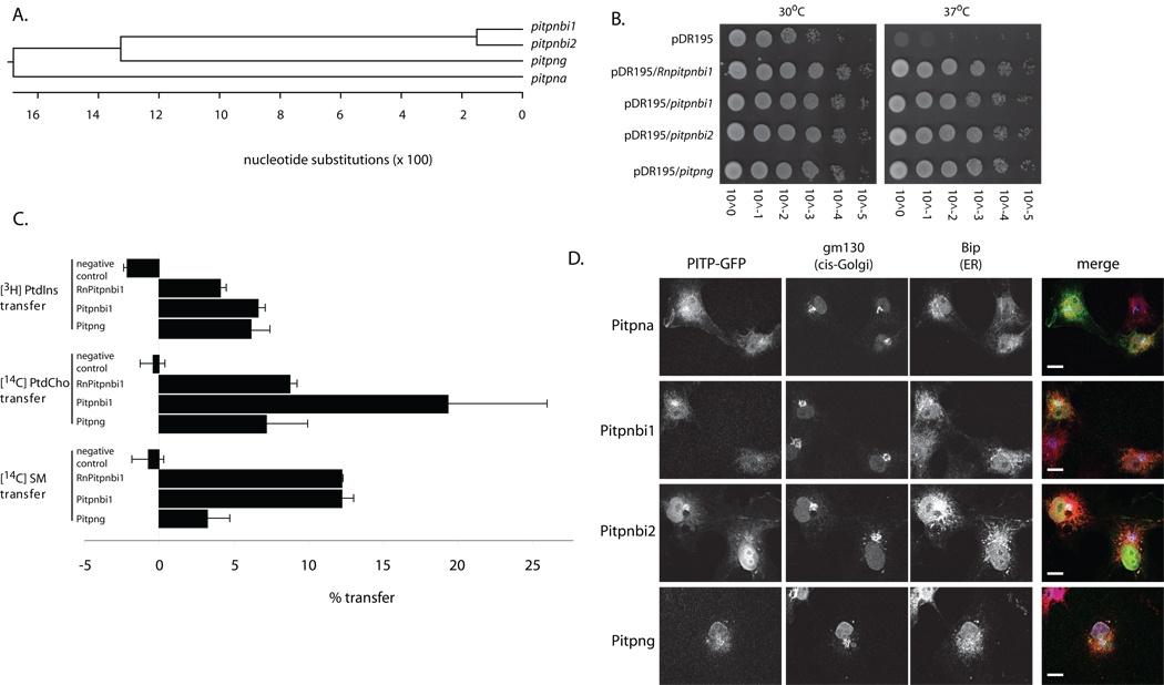
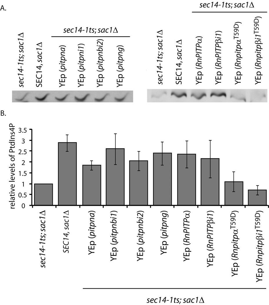
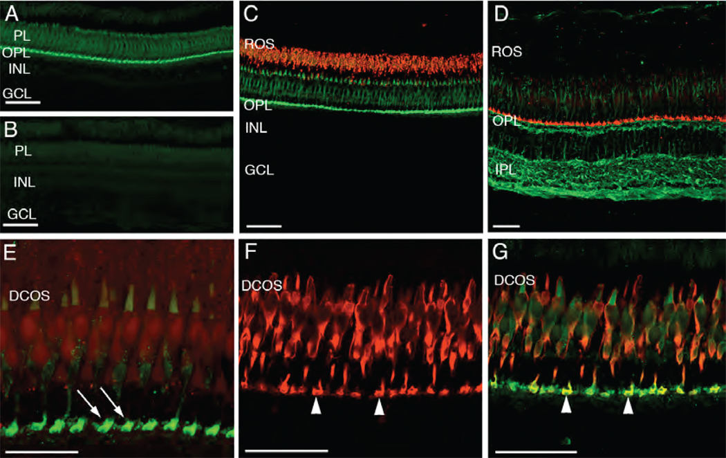
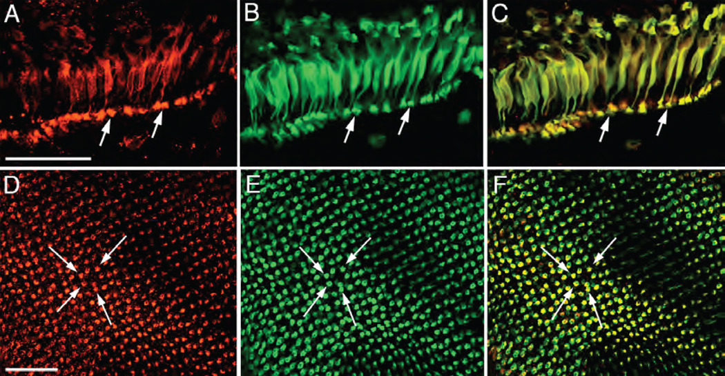
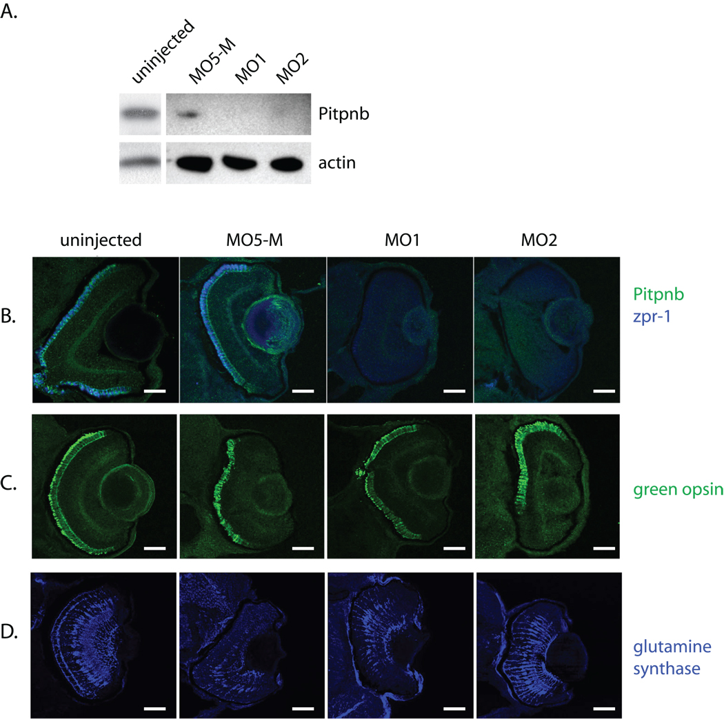
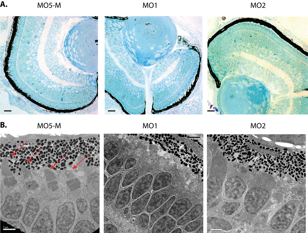
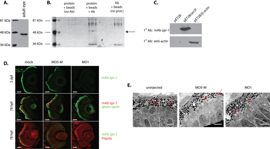
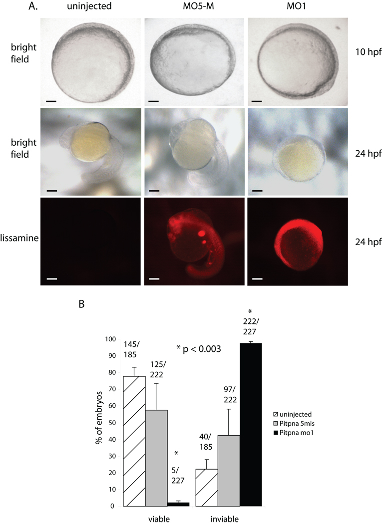
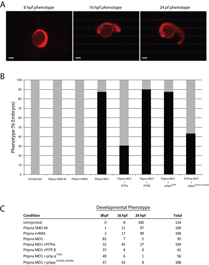
Phosphatidylinositol transfer proteins (PITPs) in yeast co-ordinate lipid metabolism with the activities of specific membrane trafficking pathways. The structurally unrelated metazoan PITPs (mPITPs), on the other hand, are an under-investigated class of proteins. It remains unclear what biological activities mPITPs discharge, and the mechanisms by which these proteins function are also not understood. The soluble class 1 mPITPs include the PITPalpha and PITPbeta isoforms. Of these, the beta-isoforms are particularly poorly characterized. Herein, we report the use of zebrafish as a model vertebrate for the study of class 1 mPITP biological function. Zebrafish express PITPalpha and PITPbeta-isoforms (Pitpna and Pitpnb, respectively) and a novel PITPbeta-like isoform (Pitpng). Pitpnb expression is particularly robust in double cone cells of the zebrafish retina. Morpholino-mediated protein knockdown experiments demonstrate Pitpnb activity is primarily required for biogenesis/maintenance of the double cone photoreceptor cell outer segments in the developing retina. By contrast, Pitpna activity is essential for successful navigation of early developmental programs. This study reports the initial description of the zebrafish class 1 mPITP family, and the first analysis of PITPbeta function in a vertebrate.


Figures

Fig. 1
Fig. 1
Pitpnb and Pitpng isoforms. (A) A ClustalW phylogenetic grouping of presumptive zebrafish Pitpna, Pitpnbi1, Pitpnbi2, and Pitpng is shown. (B) Zebrafish PITP cDNAs were subcloned into the multicopy yeast URA3 expression vector pDR195 where heterologous PITP gene expression is driven by a powerful constitutive promoter. Each individual expression construct was transformed into the yeast strain CTY-1-1A (ura3-52, sec14-1ts). Ten-fold dilution series were prepared from saturated liquid cultures normalized to similar cell densities and dilution spotted onto rich YPD solid media in parallel. One set of YPD plates was incubated for 48 hours at 30° (permissive for sec14-1ts) and the other set at 37°C (restrictive for sec14-1ts). (C) In vitro lipid transfer activities of presumptive zebrafish PITPβ-like proteins. Transfer assays score mobilization of radiolabeled PtdIns, PtdCho, or SM substrate between distinct membrane bilayer systems. Clarified cytosol prepared from yeast strain CTY303 (sec14Δ cki1) expressing each protein of interest served as protein source. Cytosol from CTY303 carrying an empty vector served as a negative control, while corresponding cytosol containing RnPITPβi1 was included as a positive control. The data shown is a single transfer assay, performed in triplicate that is representative of at least three independent experiments. The phospholipid-transfer substrate inputs for these assays were: 21,799 cpm [3H]PtdIns; 13,426 cpm [14C]PtdCho; 12,645 cpm [14C]SM;. Background values (buffer control) for these respective transfer assays were 1324, 1404 and 1038 cpm. (D) Localization of zebrafish PITPβ-like isoforms in mammalian cells. COS7 cells expressing GFP-tagged versions of zebrafish PITPs were fixed, permeabilized, and stained with antibodies that detect the cis-Golgi (gm130; blue) and the ER (BiP; red). Representative profiles for GFP expression or antibody staining are shown in the left panels, and the merged images are shown in the right panel. Scale bars -- 10 µm.
Fig. 2
Fig. 2
Zebrafish and mammalian class 1 PITPs stimulate phosphoinositide synthesis in yeast. (A) Yeast strain CTY100 (sec14-1ts sac1Δ; Cleves et al., 1989) carrying the YEp(URA3) parental expression plasmid pDR195, YEp(pitpna), YEp(pitpnbi1), YEp(pitpnbi2), YEp(pitpng), pDR195(RnPITPα), pDR195(RnPITPβi1), pDR195(RnPITPαT59D), and pDR195(RnPITPβi1T58D) were radiolabeled to steady-state with [3H]-inositol. Included was an isogenic SEC14 strain as positive control (CTY244; SEC14 sac1Δ; Cleves et al., 1989). After a 3 hour shift to 37°C, phospholipids were extracted, resolved by one-dimensional thin layer chromatography (Schaaf et al., 2008). The PtdIns-4-phosphate species are shown. (B) Quantification of PtdIns-4-phosphate. The PtdIns-4-phosphate band intensities as measured by densitometry were expressed as a ratio to PtdIns intensities for purposes of sample normalization, and the ratios are plotted as relative values. The PtdIns-4-phosphate/PtdIns ratio for the sec14-1ts sac1Δ (0.106496 – indicating a ca 10:1 molar ration of PtdIns to PtdIns-4-phosphate in this strain) was set to 1.0 on the relative scale. For comparison, a sec14-1ts strain exhibited a relative value of 0.20075 -- calculated from a PtdIns-4-phosphate/PtdIns ratio of 0.021379.
Fig. 3
Fig. 3
Pitpnb immunolocalization in the adult zebrafish retina. (A) Immunolocalization of Pitpnb. An adult retina cryosection labeled with the anti-Pitpnb serum revealed Pitpnb expression is confined to the photoreceptor layer (PL) and the outer plexiform layer (OPL). (B) A negative control section labeled with pre-immune serum shows no staining. (C) A double-label experiment. Retinal sections dual-labeled for Pitpnb (green; anti-DrPITPβ antibodies) and the zpr-3 marker (red; mAb zpr-3; revealed Pitpnb is not expressed in rods. (D) A dual-label image shows staining profiles for Pitpnb (red) and α-tubulin (green). (E) A high magnification image of a Pitpnb staining profile revealing the intense signal in the photoreceptor synaptic pedicles (arrows) in the OPL. (F) Expression of the zpr-1 staining profile (visualized via the mAb zpr-1 antibody) specifically labels double cone cells. (G) A dual-label image. The merged Pitpnb (green) and zpr-1 staining profiles (red) from frozen retinal sections are shown. Arrowheads in Panels F and G point to the double-labeled cone cell synaptic pedicles. Abbreviations: PL, photoreceptor layer; INL, inner nuclear layer; GCL, ganglion cell layer; ROS, rod outer segments; OPL, outer plexiform layer; IPL, inner plexiform layer; DCOS, double cone cell outer segments. Scale bars are 50 µm in all panels except Panel E (25 µm).
Fig. 4
Fig. 4
Pitpnb localizes to double cone cells. (A–C) Pitpnb and the double cone cell-specific zpr-1 antigen co-localize in the adult retina. Dissociated double cone cells co-labeled with anti- Pitpnb serum and mAb zpr-1. Panel A shows Pitpnb localization, Panel B the zpr-1 profile, and Panel C is a merged image. Arrows identify double cone cell synaptic pedicles. (D–F) Pitpnb antibodies and mAb zpr-1 co-label synaptic pedicles. The staining profiles in retinal wholemounts at the level of cone cell synaptic pedicles; Pitpnb profile (D), mAb zpr-1 antigen (E), and the merged images (F). Arrows highlight the same individual labeled cone pedicles in each panel. There is perfect correspondence between Pitpnb and zpr-1 signals at the level of double cone cell synaptic terminals. Scale bars -- 50 µm.
Fig. 5
Fig. 5
Reduced Pitpnb expression results in loss of zpr-1-staining. A) Morpholinos (pitpnb-directed morpholinos 1 and 2, and a 5-base mismatch control version of βMO1, and the standard specific control morpholino) were injected into 1–4 cell stage embryos. Seventy eight hours after fertilization, embryos were disrupted, solubilized in sample buffer, proteins resolved by SDS-PAGE, and transferred to nitrocellulose. Blots probed with anti-Pitpnb immunoglobulin reveal that βMO1 and βMO2, but not the controls, depress Pitpnb protein levels. (B–D) Morphants were fixed at 3 dpf, embedded in OCT, and sectioned. Retinal sections containing retina were stained for (B) Pitpnb and zpr-1 antigen, (C) green opsin, or (D) glutamine synthase. Scale bars -- 100 µm.
Fig. 6
Fig. 6
Pitpnb morphants present double cone cell structural defects. (A) Five-base-mismatch control (MO5-M) and Pitpnb morphants (3dpf) were fixed and dehydrated. After embedding in Polybed 812 resin, 3µm sections were cut and stained with 1:1 methylene blue:Azure II. No gross structural defects are observed in morphant eye sections relative to control. Scale bars -- 35 µm. (B) Ultrathin sections were stained with uranyl acetate and lead citrate, and visualized by electron microscopy. Outer cone cells are clearly recognized in the control by their well-formed and distinctive outer-segments (indicated by red arrows). These structures are absent, or otherwise unrecognizable, in the morphant retinas. Scale bars -- 2 µm.
Fig. 7
Fig. 7
Identification of zpr-1 antigen as arrestin-3-like. (A) Lysate of adult zebrafish eye was fractionated by SDS-PAGE, the resolved species transferred to nitrocellulose, and zpr-1 antigen identified by immunoblotting with mAb zpr-1. (B) mAb zpr-1 antigen was immunoisolated from adult zebrafish eye lysates and resolved by SDS-PAGE. Two controls were included: sample without lysate, and sample without mAb zpr-1. The species indicated with an arrow was uniquely recovered from the complete incubation. (C) Recombinant zebrafish β-actin and Arr3L cross-reactivity with mAb zpr-1 or anti-actin immunoglobulin was tested by blotting -- as indicated. Naïve bacterial lystaes served as negative control. (D) Embryos (1–4 cell stage) injected with morpholino against Arr3L (MO1), or the corresponding 5-base Arr3L mismatch control morpholino (MO5-M), were examined at 3 dpf for Arr3L, green opsin, and Pitpnb staining. Uninjected (mock) served as additional control. Scale bars -- 100 µm. (E) Arr3L morpholino-injected (MO1) and uninjected control embryos were fixed and prepared for ultra-thin section electron microscopy. Sections from at least 3 fish were examined and representative images are shown. Outer segments are indicated with a red arrow. Scale bars -- 2 µm.
Fig. 8
Fig. 8
Pitpna morphants fail at an early stage of development. (A) Zebrafish embryos were either injected with pitpna-directed (pitpna MO1) or the corresponding 5-base mismatch control (pitpna MO5-M) morpholinos at the 1–4 cell stage, and brightfield images were taken at the indicated time (hpf). At 24 hpf, images were also taken in the red fluorescence channel to identify embryos containing the lissamine-tagged morpholino. Top row: Scale bars -- 140 µm. Middle and bottom rows: Scale bars -- 210 µm. (B) At 48 hours post-fertilization, control and morphant embryos were classified as viable or inviable based on visual inspection. Data are from three independent experiments, and p-values are indicated.
Fig. 9
Fig. 9
Rescue of Pitpna morphant arrest phenotype by rat PITPα. (A) Wild-type 24-,16- and 8 hpf embryos injected at the 1–4 cell stage with a lissamine-tagged Standard Control morpholino, which is not complementary to any known sequence in the zebrafish genome, are shown. The red fluorescence reveals the distribution of the morpholino throughout the embryo, and these developmental phenotypes provide the phenotypic definitions for scoring rescue. Scale bars – 270 µm. (B) Wild-type embryos were either uninjected or injected with an anti-pitpna morpholino containing 5-mismatched bases (Pitpna MO5-M), in vitro transcribed rat PITPα mRNA (PITPa RNA), anti-pitpna morpholino (Pitpna MO1), anti-pitpna morpholino and rat PITPα mRNA (Pitpna MO1 + PITPb RNA), anti-pitpna morpholino and rat PITPα mRNA (Pitpna MO1 + PITPa mRNA), anti-pitpna morpholino and rat pitpW203A, W204A RNA (Pitpna MO1 + PitpaW203A,W204A mRNA), and anti-pitpna morpholino and rat PitpT59D RNA (Pitpna MO1 + PitpaT59D RNA). Preparation of capped mRNAs is described in Suppl. Materials and Methods. Duplicate experiments were performed with each injection mixture and the data were combined from the two independent injection regimes. Gray bars represent the percentage of embryos that developed at least to the 16 hours hpf phenotype by 24 hours after each treatment. The black bars correspond to the percentage of embryos that arrested at 8 hpf developmental stage at 24 hours after each treatment. (C) The total results for each condition in (B), and from which Fig 9B was generated, are tabulated according to the developmental stage achieved by 24 hours after each treatment.
Fig. 10
Fig. 10
Pitpnb function in double cone cell outer segment biogenesis and maintenance. (I) Pitpnb cooperates with a PtdIns 3-OH kinase to promote PtdIns-3-phosphate production on the outer segment. PtdIns-3-phosphate serves as a landmark on the outer segment membrane that facilitates SARA-regulated docking and fusion of opsin-containing vesicles to the outer segment (acceptor compartment). (II) Pitpnb cooperates with what would likely be a PtdIns 4-OH kinase, at the level of the double cone cell Golgi complex, to promote formation of opsin-containing vesicles that will ultimately fuse to outer segment discs in a SARA-regulated manner. (III) Pitpnb regulates signaling in the synaptic pedicle and the robustness of this signaling is transduced from the synaptic pedicle to outer segments. In this model, Pitpnb may promote formation/fusion of synaptic vesicles and, in the absence of proper synaptic signaling, the outer segment of the cone cell is not maintained. In all three scenarios, Arr3L protein stability is suggested to be dependent on its association with functional outer segments.

Similar articles

Cited by

References

    1. Berridge MJ, Irvine RF. Inositol phosphates and cell signalling. Nature. 1989;341:197–205. - PubMed
    1. Fruman DA, Meyers RE, Cantley LC. Phosphoinositide kinases. Annu Rev Biochem. 1998;67:481–507. - PubMed
    1. Strahl T, Thorner J. Synthesis and function of membrane phosphoinositides in budding yeast, Saccharomyces cerevisiae. Biochim Biophys Acta. 2007;1771:353–404. - PMC - PubMed
    1. Majerus PW. Inositol phosphate biochemistry. Annu Rev. Biochem. 1992;61:225–250. - PubMed
    1. Nishizuka Y. Protein kinase C and lipid signaling for sustained cellular responses. FASEB J. 1995;9:484–496. - PubMed

MeSH terms

Substances