Hepatitis B Virus Genotype G forms core-like particles with unique structural properties
- PMID: 20546498
- PMCID: PMC3116152
- DOI: 10.1111/j.1365-2893.2010.01330.x
Hepatitis B Virus Genotype G forms core-like particles with unique structural properties
Abstract
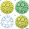
We have determined the structure of the core capsid of an unusual variant of hepatitis B virus, genotype G (HBV/G) at 14Å resolution, using cryo-electron microscopy. The structure reveals surface features not present in the prototype HBV/A genotype. HBV/G is novel in that it has a unique 36-bp insertion downstream of the core gene start codon. This results in a twelve amino acid insertion at the N-terminal end of the core protein, and two stop codons in the precore region that prevent the expression of HBeAg. HBV/G replication in patients is associated with co-infection with another genotype of HBV, suggesting that HBV/G may have reduced replication efficiency in vivo. We localized the N-terminal insertion in HBV/G and show that it forms two additional masses on the core surface adjacent to each of the dimer-spikes and have modelled the structure of the additional residues within this density. We show that the position of the insertion would not interfere with translocation of nucleic acids through the pores to the core interior compartment. However, the insertion may partially obscure several residues on the core surface that are known to play a role in envelopment and secretion of virions, or that could affect structural rearrangements that may trigger envelopment after DNA second-strand synthesis.
© 2010 Blackwell Publishing Ltd.
Figures


Similar articles
-
Hepatitis B virus nucleocapsid assembly: primary structure requirements in the core protein.J Virol. 1990 Jul;64(7):3319-30. doi: 10.1128/JVI.64.7.3319-3330.1990. J Virol. 1990. PMID: 2191149 Free PMC article.
-
Nucleic acid sequence analysis of basal core promoter/precore/core region of hepatitis B virus isolated from chronic carriers of the virus from Kolkata, eastern India: low frequency of mutation in the precore region.Intervirology. 2005;48(6):389-99. doi: 10.1159/000086066. Intervirology. 2005. PMID: 16024943
-
Nucleotide divergences in the core promoter and precore region of genotype D hepatitis B virus in patients with persistently elevated or normal ALT levels.J Clin Virol. 2001 Apr;21(1):91-101. doi: 10.1016/s1386-6532(01)00148-2. J Clin Virol. 2001. PMID: 11255102
-
Characteristics of hepatitis B virus isolates of genotype G and their phylogenetic differences from the other six genotypes (A through F).J Virol. 2002 Jun;76(12):6131-7. doi: 10.1128/jvi.76.12.6131-6137.2002. J Virol. 2002. PMID: 12021346 Free PMC article.
-
[The prevalence and clinical significance of precore and core promoter mutations in Korean patients with chronic hepatitis B virus infection].Taehan Kan Hakhoe Chi. 2002 Jun;8(2):149-56. Taehan Kan Hakhoe Chi. 2002. PMID: 12499800 Korean.
Cited by
-
Widespread hepatitis B virus genotype G (HBV-G) infection during the early years of the HIV epidemic in the Netherlands among men who have sex with men.BMC Infect Dis. 2016 Jun 10;16:268. doi: 10.1186/s12879-016-1599-7. BMC Infect Dis. 2016. PMID: 27286832 Free PMC article.
-
The scanning electron microscope in microbiology and diagnosis of infectious disease.Sci Rep. 2016 May 23;6:26516. doi: 10.1038/srep26516. Sci Rep. 2016. PMID: 27212232 Free PMC article.
-
Role of the propeptide in controlling conformation and assembly state of hepatitis B virus e-antigen.J Mol Biol. 2011 Jun 3;409(2):202-13. doi: 10.1016/j.jmb.2011.03.049. Epub 2011 Apr 2. J Mol Biol. 2011. PMID: 21463641 Free PMC article.
-
A pocket-factor-triggered conformational switch in the hepatitis B virus capsid.Proc Natl Acad Sci U S A. 2021 Apr 27;118(17):e2022464118. doi: 10.1073/pnas.2022464118. Proc Natl Acad Sci U S A. 2021. PMID: 33879615 Free PMC article.
-
Cre/LoxP-HBV plasmids generating recombinant covalently closed circular DNA genome upon transfection.Virus Res. 2021 Jan 15;292:198224. doi: 10.1016/j.virusres.2020.198224. Epub 2020 Nov 6. Virus Res. 2021. PMID: 33166564 Free PMC article.
References
-
- Steven AC, Conway JF, Cheng N, et al. Structure, assembly, and antigenicity of hepatitis B virus capsid proteins. Adv Virus Res. 2005;64:125–164. - PubMed
-
- Bottcher B, Wynne SA, Crowther RA. Determination of the fold of the core protein of hepatitis B virus by electron cryomicroscopy. Nature. 1997;386(6620):88–91. - PubMed
-
- Wynne SA, Crowther RA, Leslie AG. The crystal structure of the human hepatitis B virus capsid. Mol Cell. 1999;3(6):771–780. - PubMed
-
- Crowther RA, Kiselev NA, Bottcher B, et al. Three-dimensional structure of hepatitis B virus core particles determined by electron cryomicroscopy. Cell. 1994;77(6):943–950. - PubMed
Publication types
MeSH terms
Substances
LinkOut - more resources
Full Text Sources

