

Use of the parabiotic model in studies of cutaneous wound healing to define the participation of circulating cells
- PMID: 20546556
- PMCID: PMC2935287
- DOI: 10.1111/j.1524-475X.2010.00595.x
Use of the parabiotic model in studies of cutaneous wound healing to define the participation of circulating cells
Abstract
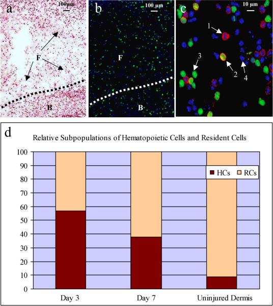
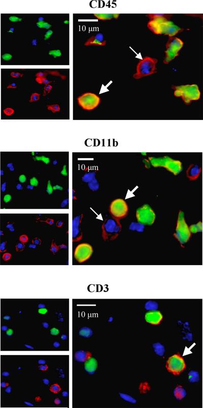
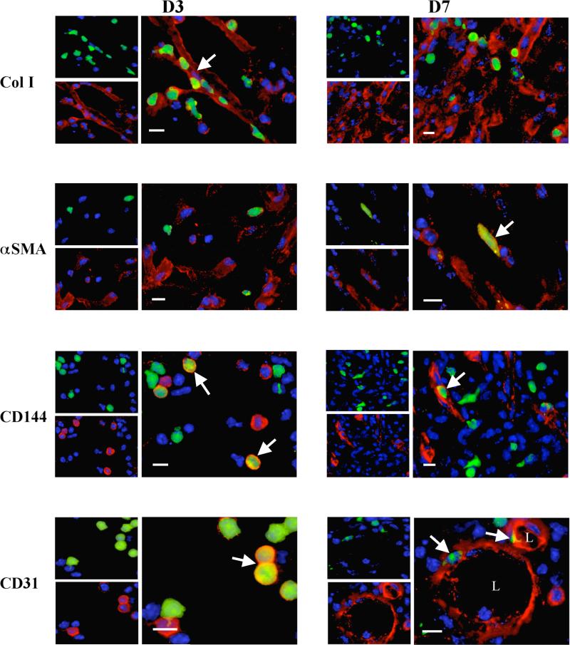
Previous experimental studies to assess the contribution of blood-borne circulating (BBC) cells to cutaneous wound healing have relied on discontinuous pulsing of labeled BBC elements or bone marrow transplant protocols. Such approaches do not allow the examination of stable BBC cells that have matured in a physiologically normal host. We have used a parabiotic murine model for cutaneous wound healing to evaluate the relative contribution of stable populations of peripheral blood cells expressing the green fluorescent protein (GFP) transgene in otherwise normal animals. Circulating cells (mature and immature) expressing the GFP transgene were easily detected and quantified in wounds of GFP- parabiotic twins during all evaluated stages of the healing response. Using multiple antibody probes, the relative contribution of various subsets of BBC cells could be comparatively assessed. In early wounds, some cells expressing mesenchymal epitopes were documented to be of hematopoietic origin, indicating the utility of this model in assessing cell plasticity in the context of tissue regeneration and repair. Application of this approach enables further investigation into the contribution of peripheral blood in normal and abnormal healing responses.
Figures
References
-
- Singer AJ, Clark RAF. Cutaneous wound healing. N Engl J Med. 1999;341(10):738–46. - PubMed
-
- Diegelmann RF, Evans MC. Wound Healing: and overview of acute, fibrotic and delayed healing. Front Biosci. 2004;9:283–9. - PubMed
-
- Fernandes KJ, McKenzie IA, Mill P, Smith KM, Akhavan M, Barnabé-Heider F, Biernaskie J, Junek A, Kobayashi NR, Toma JG, Kaplan DR, Labosky PA, Rafuse V, Hui CC, Miller FD. A dermal niche for multipotent adult skin-derived precursor cells. Nat Cell Biol. 2004;6(11):1082–93. - PubMed
-
- Cotsarelis G. Epithelial stem cells: a folliculocentric view. J Invest Dermatol. 2006;126(7):1459–68. - PubMed
-
- Opalenik SR, Davidson JM. Fibroblast differentiation of bone marrow-derived cells during wound repair. FASEB J. 2005;19(11):1561–3. - PubMed
Publication types
MeSH terms
Substances
Grants and funding
LinkOut - more resources
Full Text Sources
Other Literature Sources
Medical
