. 2010 Jun 14:10:172.
doi: 10.1186/1471-2180-10-172.

LPS-induced IL-8 activation in human intestinal epithelial cells is accompanied by specific histone H3 acetylation and methylation changes

Affiliations

LPS-induced IL-8 activation in human intestinal epithelial cells is accompanied by specific histone H3 acetylation and methylation changes

Tiziana Angrisano et al. BMC Microbiol. .

Abstract

Background: The release of LPS by bacteria stimulates both immune and specific epithelial cell types to release inflammatory mediators. It is known that LPS induces the release of IL-8 by intestinal mucosal cells. Because it is now emerging that bacteria may induce alteration of epigenetic patterns in host cells, we have investigated whether LPS-induced IL-8 activation in human intestinal epithelial cells involves changes of histone modifications and/or DNA methylation at IL-8 gene regulatory region.

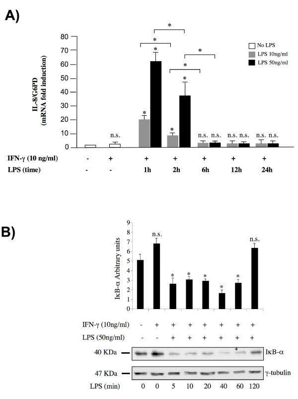
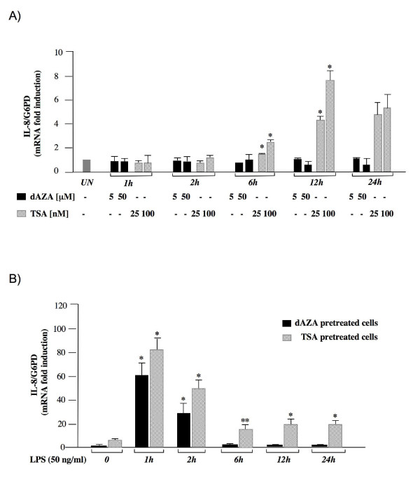
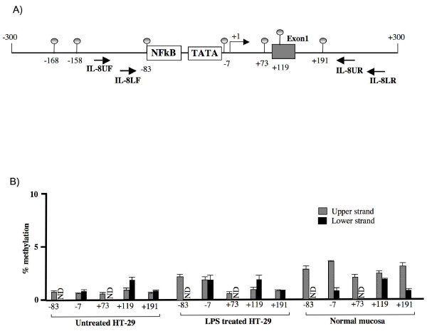
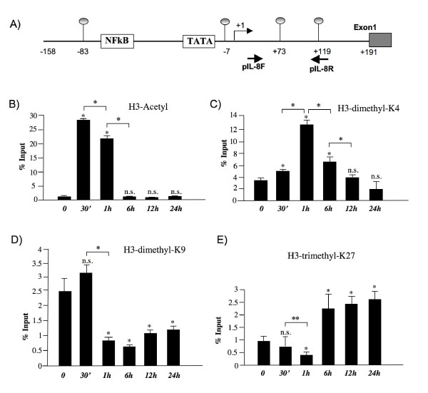
Results: RT-PCR analysis showed that IL-8 mRNA levels rapidly increase after exposure of HT-29 cells to LPS. DNA demethylating agents had no effects on IL-8 expression, suggesting that DNA methylation was not involved in IL-8 gene regulation. Consistently we found that 5 CpG sites located around IL-8 transcription start site (-83, -7, +73, +119, +191) were unmethylated on both lower and upper strand either in LPS treated or in untreated HT-29 cells, as well as in normal intestinal mucosa.Conversely, pretreatment of HT-29 cells with deacetylase inhibitors strengthened the LPS-mediated IL-8 activation. Inhibitors of histone deacetylases could induce IL-8 mRNA expression also in the absence of LPS, suggesting that chromatin modifications could be involved in IL-8 gene regulation. Chromatin immunoprecipitation analyses showed that, concurrently with IL-8 activation, transient specific changes in H3 acetylation and H3K4, H3K9 and H3K27 methylation occurred at IL-8 gene promoter during LPS stimulation. Changes of H3-acetyl, H3K4me2 and H3K9me2 levels occurred early, transiently and corresponded to transcriptional activity, while changes of H3K27me3 levels at IL-8 gene occurred later and were long lasting.

Conclusion: The results showed that specific chromatin modifications occurring at IL-8 gene, including histone H3 acetylation and methylation, mark LPS-mediated IL-8 activation in intestinal epithelial cells while it is unlikely that DNA methylation of IL-8 promoter is directly involved in IL-8 gene regulation in these cells.


Figures

Figure 1
Figure 1
Time course analysis of LPS-mediated IL-8 gene activation. (A) Total RNA was isolated at indicated time points after LPS administration and used in real-time PCR reactions. Where indicated, HT-29 cells were pre-treated with IFN-γ (10 ng/ml) for 24 hours. The IL-8 mRNA levels were normalized to G6PD levels and expressed as relative to untreated control cells. Data points represent the average of triplicate determinations ± SD. Similar results were obtained in 3 independent experiments. *, p < 0.01; n.s.= not significant, in comparison to control culture without IFN-γ. (B) Lysates were collected at the indicated time points in RIPA buffer and 50 μg of protein samples were loaded for electrophoresis. The expression levels of IκB-α were detected using anti-IκB-α antibodies. The levels of γ-tubulin were used to demonstrate equal loading. Protein levels were quantified using the software Quantity One (Bio-Rad). The IκB-α protein levels were normalized to γ-tubulin levels and expressed as relative to untreated control cells. Data points represent the average of three independent experiments ± SD. A representative blot is shown. *, p < 0.01.
Figure 2
Figure 2
Effects of TSA and 5-dAZA on IL-8 gene expression. (A) HT-29 cells were treated with the indicated concentration of drugs for the indicated times and total RNA was analyzed by real-time PCR. (B) HT-29 cells were pre-treated with 5-dAZA (50 μM) (black bars) or TSA (100 nM) (striped bars) for 24 hours (control) and then treated with LPS (50 ng/ml) and total RNA was extracted at the indicated time points after LPS administration and subjected to real time PCR analysis. The IL-8 mRNA levels were normalized to G6PD levels and expressed as relative to untreated control cells. Data points represent the average of triplicate determinations ± SD. Similar results were obtained in 3 independent experiments. Statistical analyses were performed compared to respective untreated control cells. *, p < 0.01; **, p < 0.05; absence of asterisks = not significant.
Figure 3
Figure 3
Methylation status of individual CpG sites on upper and lower strands at IL-8 promoter. (A) Schematic representation of human IL-8 gene promoter. Regulatory upstream region (proximal NF-κB binding site and TATA box), Transcriptional start site (arrow) and exon 1 (gray box) are indicated. The relative positions of each CpG site present in the analyzed region and of the primers utilized for amplification are indicated. (B) Methylation degree at CpG sites -83, -7, +73, +119, and +191 on both upper (gray bars) and lower strand (black bars) was measured in untreated HT-29, in cells treated 24 hours with LPS and in normal colon mucosa samples by MALDI-TOF analysis. Methylation of sites -83 and -73 on lower strand could not be determined by MALDI analysis (ND). Each experiment was repeated three times on three different samples. Shown are the average values for each indicated CpG site ± SD.
Figure 4
Figure 4
LPS induces early histone H3 methylation and acetylation changes at the promoter region of IL-8 gene. Chromatin from HT-29 cells was harvested at the indicated time points after LPS exposure. (A) Schematic representation of IL-8 promoter region, as in Figure 3. Positions of the primers used for ChIP analyses are shown. Presented are the results of ChIP analyses using anti-acetyl-H3 (B) anti-dimethyl-H3K4 (C), anti-dimethyl-H3K9 (D) and anti-trimethyl-K27 (E) antibodies. DNA sequences recovered after the indicated times of LPS treatment were quantified by real-time PCR using the primers indicated above. Average% input ± S.D from 4 independent experiments are plotted. *, p < 0.01; **, p < 0.05; n.s.= not significant, compared to control cells.

References

    1. Hamon MA, Cossart P. Histone modifications and chromatin remodeling during bacterial infections. Cell Host Microbe. 2008;4:100–109. doi: 10.1016/j.chom.2008.07.009. - DOI - PubMed
    1. Minárovits J. Microbe-induced epigenetic alterations in host cells: the coming era of patho-epigenetics of microbial infections. A review. Acta Microbiol Immunol Hung. 2009;56:1–19. doi: 10.1556/AMicr.56.2009.1.1. - DOI - PubMed
    1. Kouzarides T. Chromatin modifications and their function. Cell. 2007;128:693–705. doi: 10.1016/j.cell.2007.02.005. - DOI - PubMed
    1. Shilatifard A. Chromatin modifications by methylation and ubiquitination: implications in the regulation of gene expression. Annu Rev Biochem. 2006;75:243–269. doi: 10.1146/annurev.biochem.75.103004.142422. - DOI - PubMed
    1. Klose RJ, Bird AP. Genomic DNA methylation: the mark and its mediators. Trends Biochem Sci. 2006;31:89–97. doi: 10.1016/j.tibs.2005.12.008. - DOI - PubMed

Publication types

LinkOut - more resources