

Divergent signature motifs of nucleotide binding domains of ABC multidrug transporter, CaCdr1p of pathogenic Candida albicans, are functionally asymmetric and noninterchangeable
- PMID: 20546701
- PMCID: PMC2917344
- DOI: 10.1016/j.bbamem.2010.05.017
Divergent signature motifs of nucleotide binding domains of ABC multidrug transporter, CaCdr1p of pathogenic Candida albicans, are functionally asymmetric and noninterchangeable
Abstract
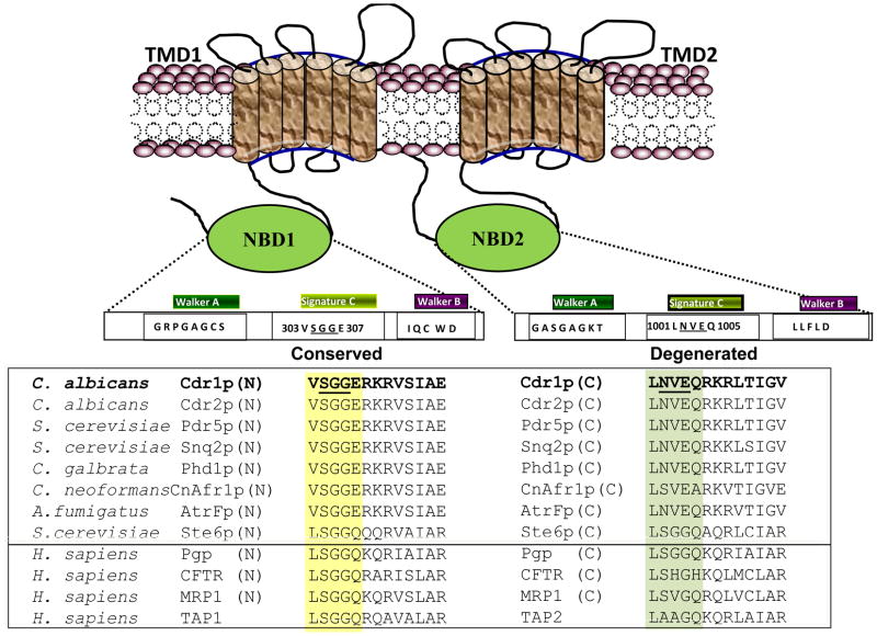
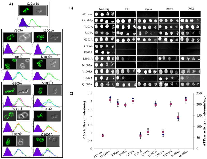
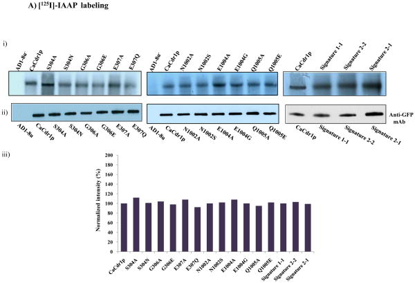
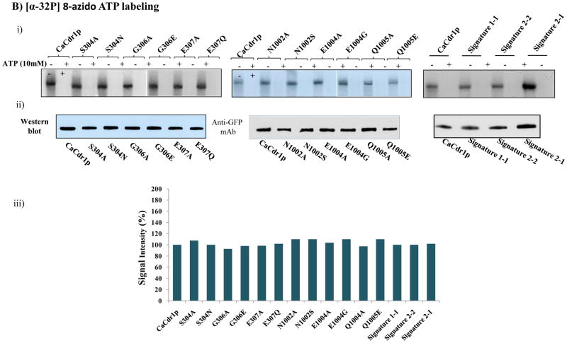
Nucleotide binding domains (NBDs) of the multidrug transporter of Candida albicans, CaCdr1p, possess unique divergent amino acids in their conserved motifs. For example, NBD1 (N-terminal-NBD) possesses conserved signature motifs, while the same motif is divergent in NBD2 (C-terminal-NBD). In this study, we have evaluated the contribution of these conserved and divergent signature motifs of CaCdr1p in ATP catalysis and drug transport. By employing site-directed mutagenesis, we made three categories of mutant variants. These included mutants where all the signature motif residues were replaced with either alanines or mutants with exchanged equipositional residues to mimic the conservancy and degeneracy in opposite domain. In addition, a set of mutants where signature motifs were swapped to have variants with either both the conserved or degenerated entire signature motif. We observed that conserved and equipositional residues of NBD1 and NBD2 and swapped signature motif mutants showed high susceptibility to all the tested drugs with simultaneous abrogation in ATPase and R6G efflux activities. However, some of the mutants displayed a selective increase in susceptibility to the drugs. Notably, none of the mutant variants and WT-CaCdr1p showed any difference in drug and nucleotide binding. Our mutational analyses show not only that certain conserved residues of NBD1 signature sequence (S304, G306, and E307) are important in ATP hydrolysis and R6G efflux but also that a few divergent residues (N1002 and E1004) of NBD2 signature motif have evolved to be functionally relevant and are not interchangeable. Taken together, our data suggest that the signature motifs of CaCdr1p, whether it is divergent or conserved, are nonexchangeable and are functionally critical for ATP hydrolysis.
2010 Elsevier B.V. All rights reserved.
Figures
 ) [34, 37].
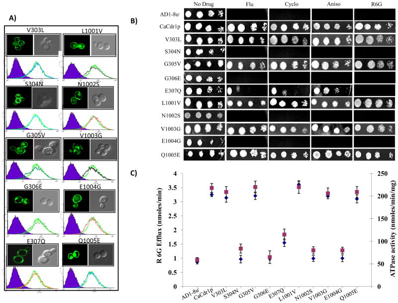 ) [34, 37].
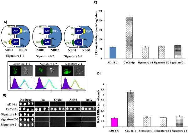
References
-
- Holland I, Cole P, Kuchler K, Higgins C. ABC Proteins from Bacteria to Man. Academic Press; San Diego, CA: 2003.
-
- Calderone RA. Candida and Candidiasis. ASM Press; Washington, DC: 2002.
-
- Prasad R, Panwar SL. Smriti, Drug resistance in yeasts—an emerging scenario. In: Poole RK, editor. Adv Microb Physiol. 1. Academic Press; London: pp. 155–201. - PubMed
Publication types
MeSH terms
Substances
Grants and funding
LinkOut - more resources
Full Text Sources
