

Genetic labeling does not detect epithelial-to-mesenchymal transition of cholangiocytes in liver fibrosis in mice
- PMID: 20546735
- PMCID: PMC2930026
- DOI: 10.1053/j.gastro.2010.05.005
Genetic labeling does not detect epithelial-to-mesenchymal transition of cholangiocytes in liver fibrosis in mice
Abstract
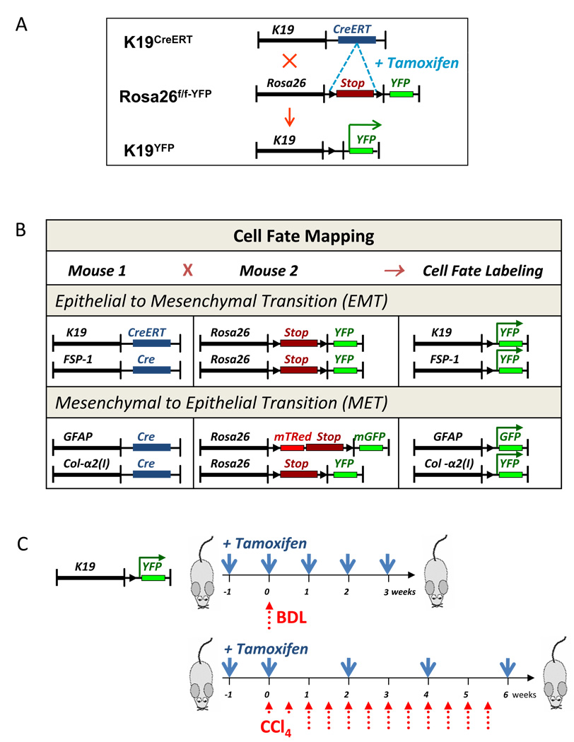
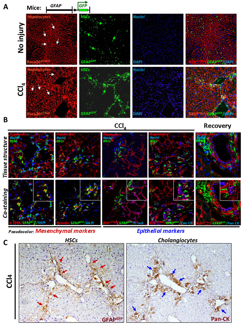
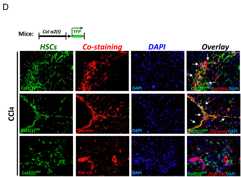
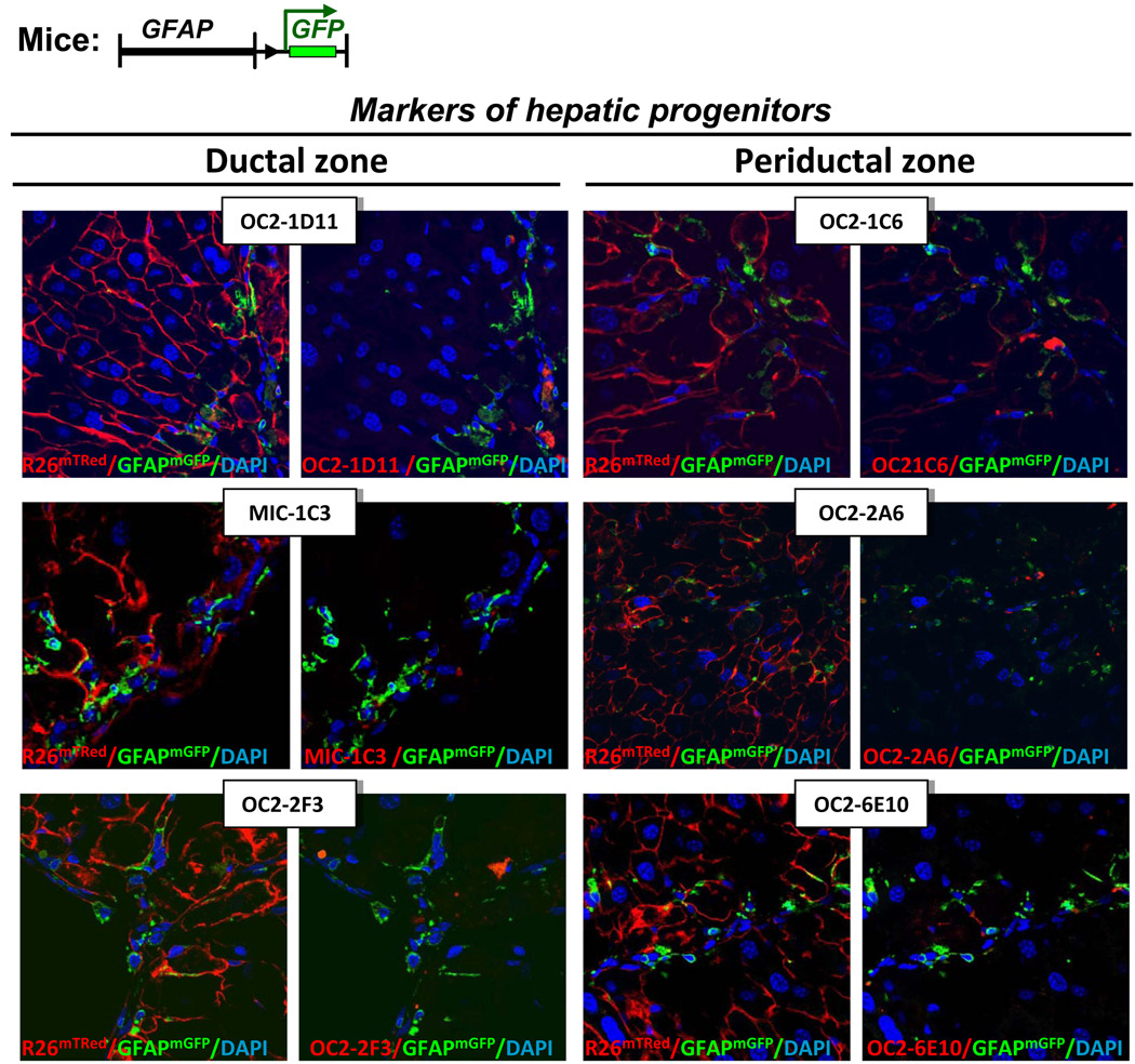
Background & aims: Chronic injury changes the fate of certain cellular populations, inducing epithelial cells to generate fibroblasts by epithelial-to-mesenchymal transition (EMT) and mesenchymal cells to generate epithelial cells by mesenchymal-to-epithelial transition (MET). Although contribution of EMT/MET to embryogenesis, renal fibrosis, and lung fibrosis is well documented, role of EMT/MET in liver fibrosis is unclear. We determined whether cytokeratin-19 positive (K19(+)) cholangiocytes give rise to myofibroblasts (EMT) and/or whether glial fibrillary acidic protein positive (GFAP(+)) hepatic stellate cells (HSCs) can express epithelial markers (MET) in response to experimental liver injury.
Methods: EMT was studied with Cre-loxP system to map cell fate of K19(+) cholangiocytes in K19(YFP) or fibroblast-specific protein-1 (FSP-1)(YFP) mice, generated by crossing tamoxifen-inducible K19(CreERT) mice or FSP-1(Cre) mice with Rosa26(f/f-YFP) mice. MET of GFAP(+) HSCs was studied in GFAP(GFP) mice. Mice were subjected to bile duct ligation or CCl(4)-liver injury, and livers were analyzed for expression of mesodermal and epithelial markers.
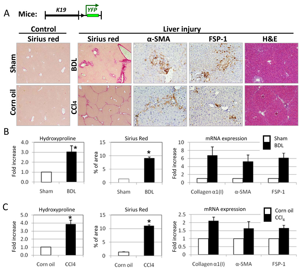
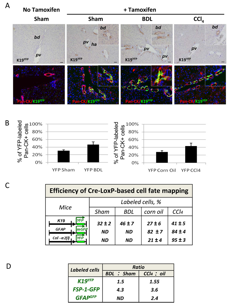
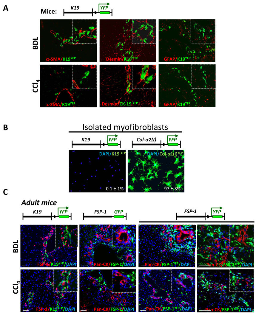
Results: On Cre-loxP recombination, >40% of genetically labeled K19(+) cholangiocytes expressed yellow fluorescent protein (YFP). All mice developed liver fibrosis. However, specific immunostaining of K19(YFP) cholangiocytes showed no expression of EMT markers alpha-smooth muscle actin, desmin, or FSP-1. Moreover, cells genetically labeled by FSP-1(YFP) expression did not coexpress cholangiocyte markers K19 or E-cadherin. Genetically labeled GFAP(GFP) HSCs did not express epithelial or liver progenitor markers in response to liver injury.
Conclusion: EMT of cholangiocytes identified by genetic labeling does not contribute to hepatic fibrosis in mice. Likewise, GFAP(Cre)-labeled HSCs showed no coexpression of epithelial markers, providing no evidence for MET in HSCs in response to fibrogenic liver injury.
Copyright © 2010 AGA Institute. Published by Elsevier Inc. All rights reserved.
Conflict of interest statement
Disclosures – the authors have no conflict of interest.
Figures
Comment in
-
Epithelial-to-mesenchymal transition in liver fibrosis: dead or alive?Gastroenterology. 2010 Sep;139(3):722-5. doi: 10.1053/j.gastro.2010.07.015. Epub 2010 Aug 1. Gastroenterology. 2010. PMID: 20682361 No abstract available.
References
-
- Parola M, Marra F, Pinzani M. Myofibroblast - like cells and liver fibrogenesis: Emerging concepts in a rapidly moving scenario. Mol Aspects Med. 2008;29:58–66. - PubMed
Publication types
MeSH terms
Substances
Grants and funding
LinkOut - more resources
Full Text Sources
Other Literature Sources
Medical
Molecular Biology Databases
Research Materials
Miscellaneous
