. 2010 Sep;139(3):1030-40.
doi: 10.1053/j.gastro.2010.05.007. Epub 2010 Jun 25.

CD39/ENTPD1 expression by CD4+Foxp3+ regulatory T cells promotes hepatic metastatic tumor growth in mice

Affiliations

CD39/ENTPD1 expression by CD4+Foxp3+ regulatory T cells promotes hepatic metastatic tumor growth in mice

Xiaofeng Sun et al. Gastroenterology. 2010 Sep.

Abstract

Background & aims: Adenosine mediates immune suppression and is generated by the ectonucleotidases CD39 (ENTPD1) and CD73 that are expressed on vascular endothelial cells and regulatory T cells (Tregs). Although tumor-infiltrating immune cells include Foxp3(+) Tregs, it is not clear whether local adenosine generation by Tregs promotes tumor growth in a CD39-dependent manner. In this study, we have examined the effect of CD39 expression by Tregs on effector immune cell responses to hepatic metastases in vivo.

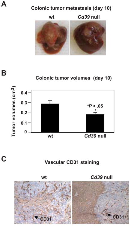
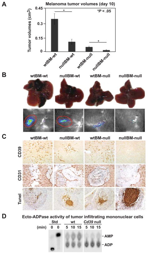
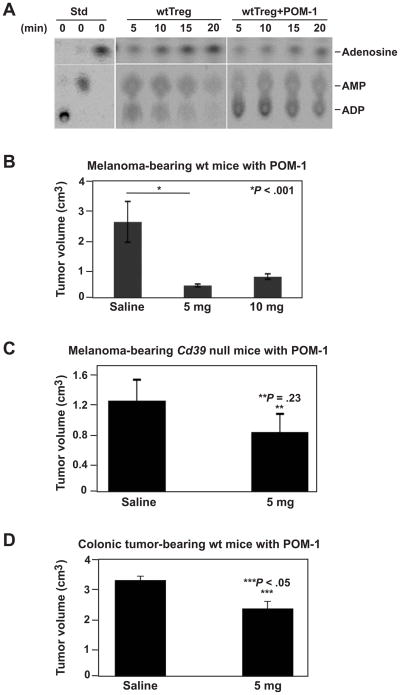
Methods: A model of hepatic metastatic cancer was developed with portal vein infusion of luciferase-expressing melanoma B16/F10 cells and MCA38 colon cancer cells in wild-type (wt) and mutant mice null for Cd39. Chimeric mice were generated by bone marrow transplantation (BMT) using Cd39 null or wt C57BL6 donors and irradiated recipient mice.

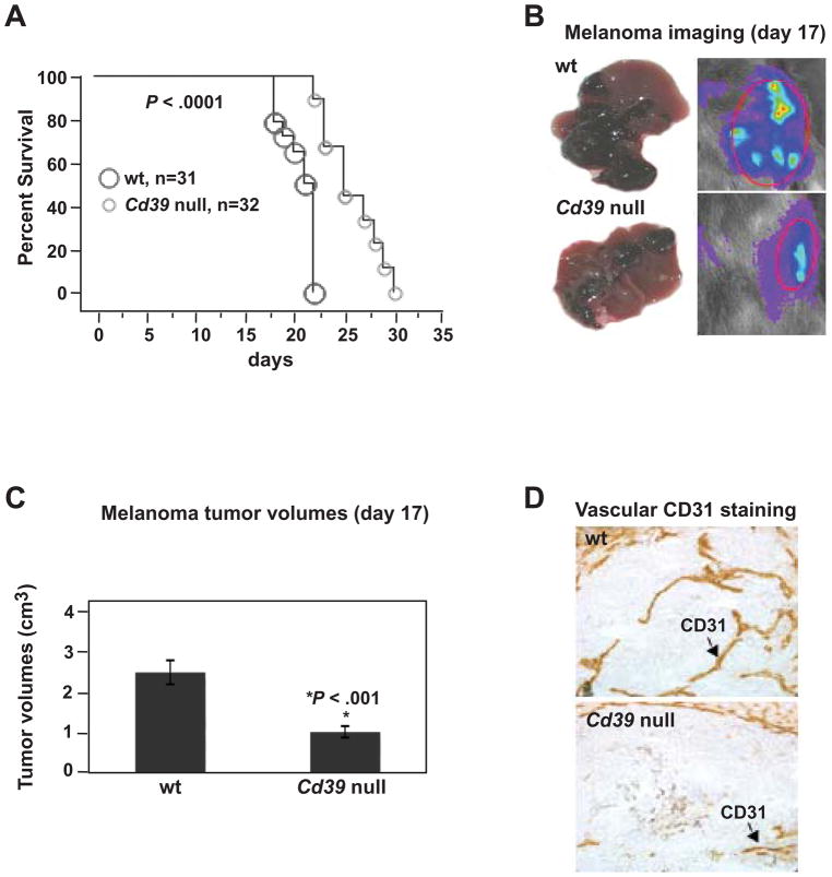
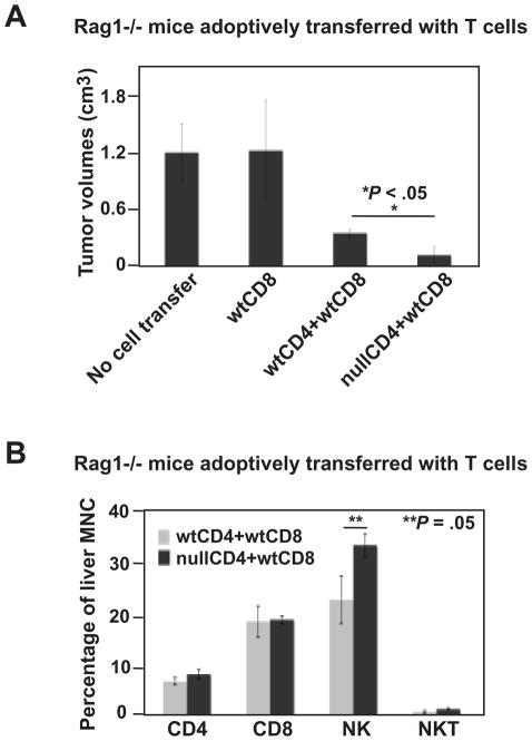
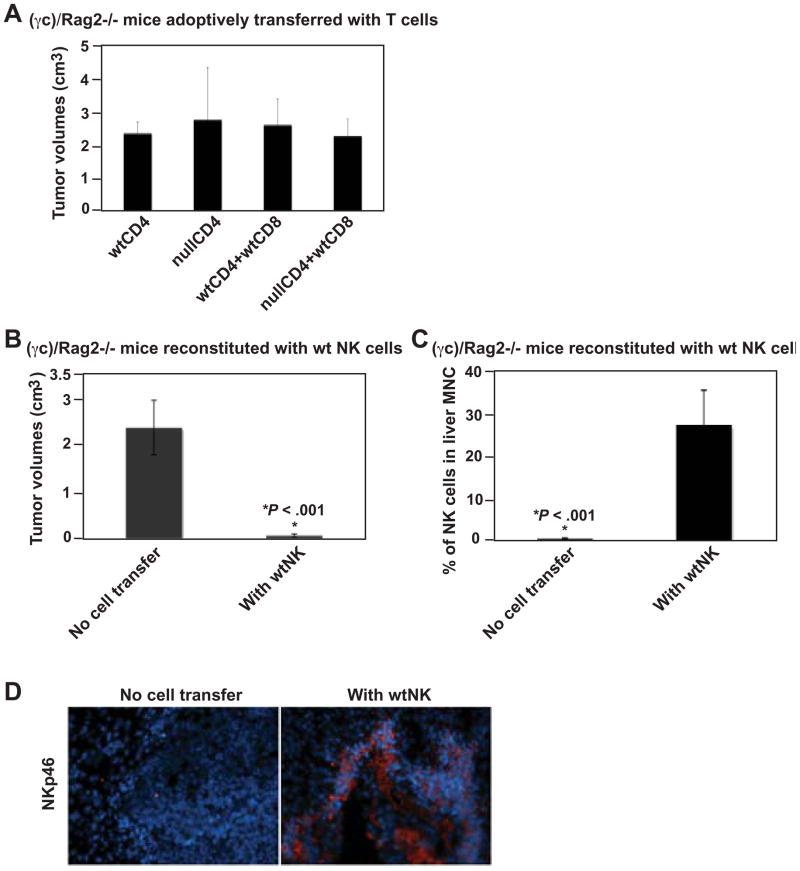
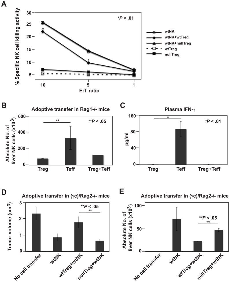
Results: We demonstrate that hepatic growth of melanoma metastatic tumors was strongly inhibited in mice with Cd39 null vasculature or in wt mice with circulating Cd39 null bone marrow-derived cells. We show functional CD39 expression on CD4(+)Foxp3(+) Tregs suppressed antitumor immunity mediated by natural killer (NK) cells in vitro and in vivo. Finally, inhibition of CD39 activity by polyoxometalate-1, a pharmacologic inhibitor of nucleoside triphosphate diphosphohydrolase activity, significantly inhibited tumor growth (P < .001).

Conclusions: CD39 expression on Tregs inhibits NK activity and is permissive for metastatic growth. Pharmacologic or targeted inhibition of CD39 enzymatic activity may find utility as an adjunct therapy for secondary hepatic malignancies.


Conflict of interest statement

Conflicts of interest: The authors disclose no conflicts.

Figures

Figure 1
Figure 1
Cd39 deletion inhibits metastatic melanoma growth in livers. (A) Survival and/or time to euthanasia in tumor-bearing mice post luc-B16/F10 cell infusion (1.5 × 10⁵ cells) via portal vein (P < .0001). (B) Representative images of tumor-bearing livers on day 17 (left panel) and in vivo bioluminescent imaging of tumor metastasis (right panel). (C) Tumor volumes at day 17 (*P < .001). (D) Representative immunohistochemical staining on tissue sections obtained from (B) using anti-CD31 (a marker for endothelium) antibody (Magnification x400). Data are given as means ± SEM.
Figure 2
Figure 2
Cd39 deletion inhibits metastatic colon tumor growth in livers. (A) Representative images of tumor-bearing livers at day 10 post portal vein infusion of 1.0 × 10⁵ MCA38 cells. (B) Tumor volumes at day 10 (*P < .05). (C) Immunohistochemical staining on tissue sections obtained from (A) using anti-CD31 (a marker for endothelium) antibody (Magnification x400). Data are given as means ± SEM.
Figure 3
Figure 3
CD39 expression on bone marrow-derived cells facilitates tumor growth. Wild type (wt) and Cd39 null (null) chimeric mice were generated by bone marrow transplantation (BMT) (donor genotype designated first/recipients second). Eight weeks post-BMT, 1.5 × 10⁵ luc-B16/F10 cells were infused via portal vein. (A) Tumor volumes on day 10 (n = 6–8 mice per group). (B) Representative images of tumor-bearing livers at day 10 (top) and in vivo bioluminescent imaging of tumor metastasis (bottom). (C) Representative immunohistochemical staining using anti-CD39 and anti-CD31 antibodies and Tunel staining (TdT) for apoptosis (Magnification x400 for CD39; x200 for CD31 and Tunel staining). (D) Ecto-ADPase activity in tumor-infiltrating mononuclear cells. Tumor-infiltrating mononuclear cells were isolated from tumor tissues of wt and Cd39 null tumor-bearing mice. Thin Layer Chromatography (TLC) analysis of ADP hydrolysis to AMP using 1.5 × 10⁵ cells was analyzed. Data are given as means ± SEM. *P < .05.
Figure 4
Figure 4
Cd39 null CD4+ T cells inhibit tumor growth. (A) Tumor volumes of tumor-bearing Rag1−/− mice adoptively transferred with lymphocytes (CD4+ T cells, 1 × 10⁶; CD8+ T cells, 0.5 × 10⁶), followed by portal vein infusion of 2 × 10⁵ luc-B16/F10 cells, on day 14. (B) Percent CD4+, CD8+, NK1.1+ and NKT lymphocytes of liver mononuclear cells isolated from tumor-bearing livers above. Data are given as means ± SEM. *P < .05, **P = .05.
Figure 5
Figure 5
NK cells mediate anti-tumor immunity to melanoma. (A) Tumor volumes of tumor-bearing (γc)/Rag2−/− mice adoptively transferred with lymphocytes (CD4+ T cells, 1 × 10⁶; CD8+ T cells, 0.5 × 10⁶), followed by portal vein infusion of 2 × 10⁵ luc-B16/F10 cells, on day 14. (B) Tumor volumes of tumor-bearing (γc)/Rag2−/− mice reconstituted with 1.5 × 10⁶ of wild type, spleen- and lymph node (LN)-derived NK cells (sorted as NK1.1+TCRβ using MOFLO), on day 14. (C) Percentage of NK cells among mononuclear cells isolated from tumor-bearing livers of (B). (D) Immunofluorescence staining using anti-NKp46 antibody on livers of (B): DAPI staining nuclei in blue and NKp46 in red (Magnification x200). Data are given as means ± SEM. *P < .001.
Figure 6
Figure 6
Treg regulate NK cell-mediated anti-tumor immunity in a CD39-dependent manner. (A) Cd39 null Treg fail to suppress NK cell cytotoxicity in vitro. NK cells were cocultured at 1.5:1 and other ratios (not shown) with activated wt or Cd39 null Treg. The sorted CD4+GFP+ Treg were stimulated with anti-CD3/CD28 bound MACSiBead Particles and IL2 (100 ng/ml) for 72 h before coculture. (*P < .01, wtNK vs wtNK+wtTreg). (B) Absolute liver NK cell numbers in tumor-bearing Rag1−/− mice adoptively transferred with sorted wt lymphocytes (Treg, 0.1 × 10⁶; Teff, 0.9 × 10⁶) as indicated, followed by portal vein infusion of 2 × 10⁵ luc-B16/F10 cells, on day 14. (C) Levels of IFN-γ in plasma obtained from mice in (B). (D) Tumor volumes of tumor-bearing (γc)/Rag2−/− mice adoptively transferred with lymphocytes (Treg, 1.0 × 10⁶; NK, 1.0 × 10⁶), followed by portal vein infusion of 2 × 10⁵ luc-B16/F10 cells, on day 14. Mice received PBS as controls. (E) Absolute liver NK cell numbers in tumor-bearing mice of (D). Data are given as means ± SEM. *P < .01, **P < .05.
Figure 7
Figure 7
Pharmacological inhibition of CD39 activity suppresses tumor growth. (A) Ecto-NTPDase activity of POM-1-treated wt Treg in vitro. Wt Treg (CD39+CD4+Foxp3+, 3 × 10⁵) were pre-treated with 26 μm of POM-1, washed, and then subjected to TLC ADPase analysis. Treg incubated in POM-1-free medium served as control. (B) Tumor volumes of melanoma-bearing wt mice (received 2 × 10⁵ luc-B16/F10 cells) treated with POM-1 on a daily dosage of 5 or 10 mg/kg for 10 days, on day 14. Mice received saline were used as controls. (C) Tumor volumes of melanoma-bearing Cd39 null mice (received 2 × 10⁵ luc-B16/F10 cells) treated with POM-1 on a daily dosage of 5 mg/kg for 10 days, on day 14. Mice received saline served as controls. (D) Tumor volumes of colonic tumor-bearing wt mice (received 2 × 10⁵ MCA38 cells) treated with POM-1 on a daily dosage of 5 mg/kg for 10 days, on day 14. Mice received saline were used as controls. Data are given as means ± SEM. *P < .001, **P = .23, ***P < .05.

References

    1. Ogawa T, Takayama K, Takakura N, Kitano S, Ueno H. Anti-tumor angiogenesis therapy using soluble receptors: enhanced inhibition of tumor growth when soluble fibroblast growth factor receptor-1 is used with soluble vascular endothelial growth factor receptor. Cancer Gene Ther. 2002;9:633–40. - PubMed
    1. Chari RS, Helton WS, Marsh RD. Chemotherapy and regional therapy of hepatic colorectal metastases: expert consensus statement by Bartlett et al. Ann Surg Oncol. 2006;13:1293–5. - PubMed
    1. Hoskin DW, Reynolds T, Blay J. Adenosine as a possible inhibitor of killer T-cell activation in the microenvironment of solid tumours. Int J Cancer. 1994;59:854–5. - PubMed
    1. Burnstock G. Purinergic signaling and vascular cell proliferation and death. Arterioscler Thromb Vasc Biol. 2002;22:364–73. - PubMed
    1. Spychala J. Tumor-promoting functions of adenosine. Pharmacol Ther. 2000;87:161–73. - PubMed

Publication types

MeSH terms