Review
. 2010 Dec 30;62(15):1456-67.
doi: 10.1016/j.addr.2010.05.004. Epub 2010 May 27.

Drug delivery to solid tumors by elastin-like polypeptides

Affiliations
Review

Drug delivery to solid tumors by elastin-like polypeptides

Jonathan R McDaniel et al. Adv Drug Deliv Rev. .

Abstract

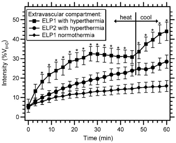
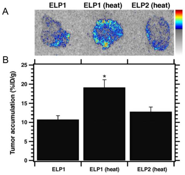
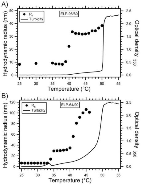
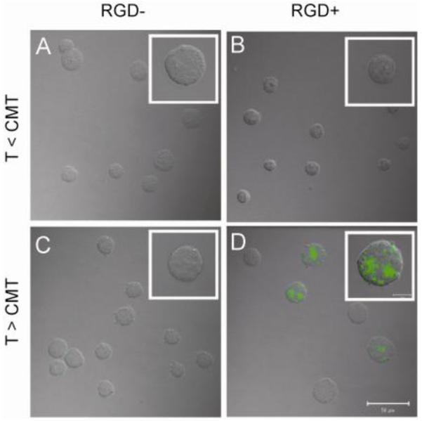
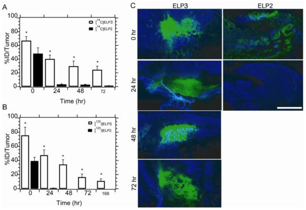
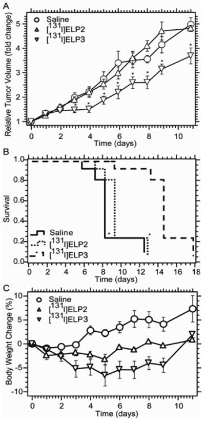
Thermally responsive elastin-like polypeptides (ELPs) are a promising class of recombinant biopolymers for the delivery of drugs and imaging agents to solid tumors via systemic or local administration. This article reviews four applications of ELPs to drug delivery, with each delivery mechanism designed to best exploit the relationship between the characteristic transition temperature (T(t)) of the ELP and body temperature (T(b)). First, when T(t)≫T(b), small hydrophobic drugs can be conjugated to the C-terminus of the ELP to impart the amphiphilicity needed to mediate the self-assembly of nanoparticles. These systemically delivered ELP-drug nanoparticles preferentially localize to the tumor site via the EPR effect, resulting in reduced toxicity and enhanced treatment efficacy. The remaining three approaches take direct advantage of the thermal responsiveness of ELPs. In the second strategy, where T(b)<T(t)<42°C, an ELP-drug conjugate can be injected in conjunction with external application of mild hyperthermia to the tumor to induce ELP coacervation and an increase in concentration within the tumor vasculature. The third approach utilizes hydrophilic-hydrophobic ELP block copolymers that have been designed to assemble into nanoparticles in response to hyperthermai due to the independent thermal transition of the hydrophobic block, thus resulting in multivalent ligand display of a ligand for spatially enhanced vascular targeting. In the final strategy, ELPs with T(t)<T(b) are conjugated with radiotherapeutics, injtect intioa tumor where they undergo coacervation to form an injectable drug depot for intratumoral delivery. These injectable coacervate ELP-radionuclide depots display a long residence in the tumor and result in inhibition of tumor growth.


Figures

Figure 1
Figure 1. ELP drug delivery strategies
This figure depicts four strategies that use ELPs to deliver drugs to a solid tumor in vivo. (A) Drug attachment triggered self-assembly of an ELP into micelles. Hydrophobic drugs are attached to the C-terminus of a hydrophilic ELP to trigger the self-assembly of micelles. These micelles accumulate in the tumor by passive diffusion through the leaky tumor vasculature. (B) Thermal targeting of an ELP to a heated tumor by the phase transition triggered aggregation of the ELP in tumor vasculature. ELP-drug conjugates can be actively targeted to a tumor by using the ELP phase transition in combination with the application of mild hyperthermia to the tumor to trigger formation of micron sized aggregates of the ELP that adhere to the vessel walls. Upon cessation of hyperthermia, the aggregates dissolve, generating a large concentration gradient that drives the ELP that dissolves from the aggregates into the tumor. (C) Multivalent targeting of a solid tumor by thermally triggered self-assembly of a diblock ELP into micelles in a heated tumor. Focused mild hyperthermia of a solid tumor can be used to thermally trigger the self-assembly of diblock ELPs into micelles that display a tumor targeting ligand on the corona of the micelle. In ELP unimers, the ligand is monovalent and hence has low avidity, which does not lead to significant ELP uptake by cells. In the thermally triggered micelle state, multivalent presentation of the ligand leads to high avidity and greater uptake by cells. (D) Local delivery of an ELP that coacervates in a solid tumor upon intratumoral injection. An ELP with a Tt below body temperature can be directly injected into a tumor to form an insoluble coacervate which forms a long-lasting depot, which extends the exposure of a conjugated radiotherapeutic to the tumor.
Figure 2
Figure 2. Characterization of CP-Dox nanoparticles
CP-Dox nanoparticles characterized by: (A) freeze fracture scanning electron microscopy (bar = 200 nm) and (B) dynamic light scattering at 25 μM in PBS at 37 °C. DLS measures the distribution of hydrodynamic radii (Rh) [27].
Figure 3
Figure 3. Plasma pharmacokinetics and tissue biodistribution
(A) Plasma concentration of Dox as a function of time post-injection. Lines represent a two-compartment model fit. Error bars indicate the 95% confidence interval (n=5). (B) Dox concentration in tumor tissue. * p < 0.0005 Analysis of Variance, Tukey's HSD. Data are presented as mean ± SD [27].
Figure 4
Figure 4. Anti-tumor activity of CP-Dox nanoparticles
(A,B) Tumor cells (C26) were implanted subcutaneously on the back of the mice on day zero. Mice were treated on day 8 (↑) at the MTD with PBS (n=10), free Dox (5 mg / kg BW; n=10), or CP-Dox (20 mg Dox equivalent/ kg BW; n=9). a) Tumor volume up to day 30 (for n > 6). Error bars represent the SEM. *indicates p= 0.03, 0.00002 for CP-Dox vs. Dox and CP-Dox vs. PBS (day 15), respectively (Mann Whitney). b) Cumulative survival of mice **indicates p = 0.0001, 0.00004 for CP-Dox vs. Dox and CP-Dox vs. PBS, respectively (Kaplan- Meier) [27].
Figure 5
Figure 5
Images of thermally sensitive ELP1 (green) and thermally insensitive ELP2 (red) in a solid tumor before, during, and after hyperthermia treatment. The vascular intensities of the ELPs in (A) were balanced to produce a yellow color. (A) 0 min, before heat application. (B) 41.5 °C, after 30 minutes of heat application. The green punctate fluorescence indicates aggregation of ELP1. (C) 37 °C, after 10 minutes of cooling. The absence of punctate fluorescence in (C) as compared to (B) demonstrates the reversibility of ELP1's phase transition. The bar represents 100 μm in all images [29].
Figure 6
Figure 6
Extravascular accumulation of a thermally sensitive ELP with hyperthermia treatment as a function of time. Data were normalized by the initial vascular intensity for each animal and expressed as a percentage of vascular intensity at t = 0 min. The tumor was not heated for the ELP1 normothermia control. The tumor was heated to 41.5 °C for the first 45 min and then cooled to 37 °C for the remaining 15 min for the ELP1 hyperthermia and ELP2 hyperthermia conditions. The data are expressed as mean ± SE. *, P<0.05, Fisher's PLSD for ELP1 with hyperthermia versus ELP2 with hyperthermia [29].
Figure 7
Figure 7
(A) Autoradiography images of 20 μm tumor sections after 1 hr of hyperthermia treatment with 14C labeled ELP1 with and without heat, and 14C labeled ELP2 with heat. (B) Scintillation analysis of the tumor sections shows that ELP1 with heat results in significantly greater accumulation than either of the controls. Data are shown as mean ± SEM. *P < 0.05, Fisher's PLSD [23].
Figure 8
Figure 8. Thermally-triggered self-assembly of ELPBC micelles
Optical density at 350 nm and hydrodynamic radius (Rh) of (A) 25 dμM ELP[VA8G7-96]/[V5-60] and (B) ELP[VA8G7-64]/[V5-90] in PBS versus temperature [30].
Figure 9
Figure 9. Thermally-triggered multivalency increases uptake of ELPBC micelles
Alexa488-labeled ELP[VA8G7-64]/[V5-90] without (A,C) and with (B,D) an N-terminal RGD ligand were incubated at 10 μM for 1 h with K562 cells expressing αvβ3 below (A,B) and above (C,D) the CMT of the ELPBC. Confocal microscopy demonstrates that significant uptake only occurred when the RGD-presenting ELPBC was incubated above its CMT (D) [64].
Figure 10
Figure 10. Tumor retention of ELP coacervate
(A) Tumor retention of 14C-ELPs following intratumoral administration to FaDu tumors implanted s.c. in Balb/c nu/nu mice. The radioactivity present in the tumor was measured using a beta-counter and expressed as the percentage of the injected dose. Data are presented as the mean, with error bars representing the SEM (n=8-10; *: p < 0.01, t-test). (B) Tumor retention of 125I-labeled ELPs following administration to 4T1 tumors as described above, measured using a gamma counter. (C) Fluorescence images of tumor sections following administration of ELPs labeled with Alexa-Fluor 488 (green), with Hoechst nuclear stain (blue) administered prior to sacrifice (scale bar = 100 μm) [31].
Figure 11
Figure 11. Antitumor efficacy of 131I-ELP coacervate
(A) Relative volume of s.c. 4T1 tumors in Balb/c nu/nu mice following intratumoral administration of 131I-ELP3, 131I-ELP2-128, and saline, presented as the mean of n = 9 with error bars representing SEM (*: p < 0.05 vs. ELP2-128 or saline). (B) Kaplan-Meier analysis of survival, with 5x initial tumor volume defined as the endpoint. (C) Change in body weight following i.t. administration of 131I-ELP3, 131I-ELP2-128, or saline, presented as the mean of n = 9 with error bars representing SEM [31].

Similar articles

Cited by

References

    1. Duncan R. Polymer conjugates as anticancer nanomedicines. Nature Reviews Cancer. 2006;6(9):688–701. - PubMed
    1. Vasey PA, Kaye SB, Morrison R, Twelves C, Wilson P, Duncan R, Thomson AH, Murray LS, Hilditch TE, Murray T, Burtles S, Fraier D, Frigerio E, Cassidy J, I.I.I.C. Canc Res Campaign Phase Phase i clinical and pharmacokinetic study of pk1 n-(2-hydroxypropyl)methacrylamide copolymer doxorubicin : First member of a new class of chemotherapeutic agents - drug-polymer conjugates. Clinical Cancer Research. 1999;5(1):83–94. - PubMed
    1. Chuang VTG, Kragh-Hansen U, Otagiri M. Pharmaceutical strategies utilizing recombinant human serum albumin. Pharmaceutical Research. 2002;19(5):569–577. - PubMed
    1. Vandamme TF, Lenourry A, Charrueau C, Chaumeil J. The use of polysaccharides to target drugs to the colon. Carbohydrate Polymers. 2002;48(3):219–231.
    1. Allen TM. Liposomes - opportunities in drug delivery. Drugs. 1997;54:8–14. - PubMed

Publication types