HIV-miR-H1 evolvability during HIV pathogenesis
- PMID: 20546828
- PMCID: PMC3478900
- DOI: 10.1016/j.biosystems.2010.05.001
HIV-miR-H1 evolvability during HIV pathogenesis
Abstract
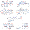
The discovery of microRNAs (miRNAs) in viruses has generated considerable attention into their functional relevance in processes such as cell death, viral proliferation, and oncogenesis. Two early studies found no detectable miRNAs expressed within HIV; however, several studies have verified the existence and function of three HIV miRNAs, most notably HIV-miR-TAR, thus making the earlier results controversial. Although miRNAs are highly conserved within most species, HIV is known to have a high mutation rate, which could contribute to the opposing experimental findings and raises questions about whether all HIV miRNAs are robust enough to maintain their integrity, especially in viral regions prone to insertions and deletions. In addition, could the evolvability of HIV miRNAs contribute to the diversity in HIV disease pathogenesis? To address this question, we examined mutations in 1293 sequences in a suspect HIV miRNA, called miR-H1, derived from a large variety of tissues from seven patients. We found considerable diversity within the structures, including a patient-specific deletion and the potential for the development of new miRNAs as a result of deletions. We also note a potential disease association between a less stable miR-H1 and the development of AIDS-related lymphoma (ARL).
Figures


Similar articles
-
Identification of human microRNA-like sequences embedded within the protein-encoding genes of the human immunodeficiency virus.PLoS One. 2013;8(3):e58586. doi: 10.1371/journal.pone.0058586. Epub 2013 Mar 8. PLoS One. 2013. PMID: 23520522 Free PMC article.
-
Genetic and biological analysis of variants derived from the cerebrospinal fluid of HIV type 1 subtype B- and D- infected patients with and without AIDS dementia complex.AIDS Res Hum Retroviruses. 1996 Nov 20;12(17):1643-5. doi: 10.1089/aid.1996.12.1643. AIDS Res Hum Retroviruses. 1996. PMID: 8947301 No abstract available.
-
Highly divergent env sequences of HIV-1 B subtype with two novel V3 loop motifs detected in an AIDS patient in Miami, Florida.AIDS Res Hum Retroviruses. 1995 Sep;11(9):1139-41. doi: 10.1089/aid.1995.11.1139. AIDS Res Hum Retroviruses. 1995. PMID: 8554913 No abstract available.
-
Transgenic mice as models of human immunodeficiency virus expression and related cellular effects.J Gen Virol. 1994 Oct;75 ( Pt 10):2549-58. doi: 10.1099/0022-1317-75-10-2549. J Gen Virol. 1994. PMID: 7931142 Review. No abstract available.
-
MICRORNA BIOGENESIS AND ITS ROLE IN HIV-1 INFECTION.Roum Arch Microbiol Immunol. 2014 Jul-Dec;73(3-4):84-91. Roum Arch Microbiol Immunol. 2014. PMID: 26201123 Review.
Cited by
-
MicroRNAs and long non-coding RNAs during transcriptional regulation and latency of HIV and HTLV.Retrovirology. 2024 Feb 29;21(1):5. doi: 10.1186/s12977-024-00637-y. Retrovirology. 2024. PMID: 38424561 Free PMC article. Review.
-
Epigenetic control of HIV-1 post integration latency: implications for therapy.Clin Epigenetics. 2015 Sep 24;7:103. doi: 10.1186/s13148-015-0137-6. eCollection 2015. Clin Epigenetics. 2015. PMID: 26405463 Free PMC article. Review.
-
Small non-coding RNAs encoded by RNA viruses: old controversies and new lessons from the COVID-19 pandemic.Front Genet. 2023 Jun 21;14:1216890. doi: 10.3389/fgene.2023.1216890. eCollection 2023. Front Genet. 2023. PMID: 37415603 Free PMC article. Review.
-
Bovine leukemia virus pre-miRNA genes' polymorphism.RNA Biol. 2018;15(12):1440-1447. doi: 10.1080/15476286.2018.1555406. RNA Biol. 2018. PMID: 30513054 Free PMC article.
-
Exosomes derived from HIV-1-infected cells contain trans-activation response element RNA.J Biol Chem. 2013 Jul 5;288(27):20014-33. doi: 10.1074/jbc.M112.438895. Epub 2013 May 9. J Biol Chem. 2013. PMID: 23661700 Free PMC article.
References
-
- Aquino-De Jesus MJ, Anders C, et al. Genetically and epidemiologically related “non-syncytium-inducing” isolates of HIV-1 display heterogeneous growth patterns in macrophages. Journal of Medical Virology. 2000;61 (2):171–180. - PubMed
-
- Bartkova J, Horejsi Z, et al. DNA damage response as a candidate anti-cancer barrier in early human tumorigenesis. Nature. 2005;434 (7035):864–870. - PubMed
-
- Briggs DR, Tuttle DL, et al. Envelope V3 amino acid sequence predicts HIV-1 phenotype (co-receptor usage and tropism for macrophages) AIDS. 2000;14 (18):2937–2939. - PubMed
Publication types
MeSH terms
Substances
Grants and funding
LinkOut - more resources
Full Text Sources
Medical

