Comparative Study
. 2010 Jun 10;66(5):663-70.
doi: 10.1016/j.neuron.2010.05.002.

Assessing spinal axon regeneration and sprouting in Nogo-, MAG-, and OMgp-deficient mice

Affiliations
Comparative Study

Assessing spinal axon regeneration and sprouting in Nogo-, MAG-, and OMgp-deficient mice

Jae K Lee et al. Neuron. .

Abstract

A central hypothesis for the limited capacity for adult central nervous system (CNS) axons to regenerate is the presence of myelin-derived axon growth inhibitors, the role of which, however, remains poorly understood. We have conducted a comprehensive genetic analysis of the three major myelin inhibitors, Nogo, MAG, and OMgp, in injury-induced axonal growth, including compensatory sprouting of uninjured axons and regeneration of injured axons. While deleting any one inhibitor in mice enhanced sprouting of corticospinal or raphespinal serotonergic axons, there was neither associated behavioral improvement nor a synergistic effect of deleting all three inhibitors. Furthermore, triple-mutant mice failed to exhibit enhanced regeneration of either axonal tract after spinal cord injury. Our data indicate that while Nogo, MAG, and OMgp may modulate axon sprouting, they do not play a central role in CNS axon regeneration failure.


Figures

Figure 1
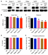
Figure 1. Basic characterization of Nogo/MAG/OMgp triple mutants
(A) Western blot analysis of Nogo-A, MAG, OMgp, NgR1 and PirB on total brain extracts from WT and Nogo/MAG/OMgp mutant mice. WT, wild type; KO, knockout (mutant); Ab, Antibody. Representative results are shown from one out of 2–3 independent biological replicates that gave similar results. (B–E) Baseline behavioral performance of WT, single and triple mutants in various locomotor tasks used in the spinal cord injury models (B–D, n = 17–24) or the forepaw preference test used in the pyramidotomy model (E, n = 8–11). *P < 0.05 compared with WT. One-way ANOVA with Tukeys post-test. All error bars are s.e.m.
Figure 2
Figure 2. Lack of a synergistic effect of deleting Nogo, MAG, and OMgp in releasing myelin inhibition in vitro
(A-E) Representative images (A–D) and quantification (E) of neurite outgrowth from WT postnatal mouse cerebellar granule neurons (CGNs) plated on laminin or CNS myelin from mice of various genotypes. (F–K) Representative images (F, G, I, J) and quantification (H, K) of neurite outgrowth from dissociated adult WT mouse dorsal root ganglion (DRG) neurons (F, G, H) or DRG explants (I, J, K) cultured on top of adult spinal cord sections of various genotypes. Max length, longest neurite length. Results are shown from one out of three experiments that gave similar results. All error bars are s.e.m. n > 120 (E); n > 30 (H); n > 25 (K). @P < 0.05 compared with Laminin control. *P < 0.05 compared with WT. One-way ANOVA with Tukeys post-test. Scale bars: 100 μm (A–D, I, J), 50 μm (F, G).
Figure 3
Figure 3. Sprouting of raphespinal serotonergic axons and locomotor recovery after lateral hemisection
(A, B) Representative images of transverse sections of lumbar spinal cord immunostained for serotonergic (5-HT) axons in a WT mouse (A) and a triple mutant (B). (C) Quantification of 5-HT immunoreactivity at the lumbar enlargement ipsilateral to the lateral hemisection (n = 9–13). *P < 0.05 compared with WT, one-way ANOVA with Tukeys post-test. (D) Gridwalk behavioral recovery. Two-way repeated measures ANOVA with Bonferonni post-test. All error bars are s.e.m. Scale bars: 500 μm. See also Figure S1.
Figure 4
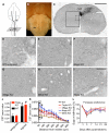
Figure 4. Sprouting of CST axons and recovery of forepaw preference after pyramidotomy
(A) Illustration of the pyramidotomy model (dorsal view). Arrow, site of pyramidotomy; shaded area, the plane of section for (C); arrowhead, axonal sprouts from the uninjured side. (B) Representative ventral view of the boxed area in (A) to show the site of pyramidotomy (arrow). (C) Representative transverse spinal cord section labeled for uninjured CST axons at the cervical enlargement following pyramidotomy. Solid rectangle represents the region quantified in (J, K). (D–I) Representative higher magnification images corresponding to the dotted area in (C) from mice of various genotypes. (J, K) Quantification of labeled uninjured CST axons at cervical levels comparing uninjured and injured mice (J), or comparing all genotypes following pyramidotomy (K). Rectangle in (K) indicates the data points depicted for injured mice in (J). Uninjured mice: n = 2–3 mice/genotype; pyramidotomized mice: n = 8–11 mice/genotype. (L) Recovery of forepaw preference. *P < 0.05 compared with WT; #P < 0.05 compared with uninjured. Two-way repeated measure ANOVA with Bonferonni post-test. All error bars are s.e.m. Scale bars: 500 μm (C), 50 μm (D–I). See also Figures S2 and S3.
Figure 5
Figure 5. Lack of regeneration of CST and raphespinal serotonergic axons in triple mutant mice
(A–D) Representative images (A, B) and quantification (C, n = 10–12) of traced CST axons in sagittal sections, and gridwalk behavioral recovery (D) following a dorsal hemisection injury. Arrows indicate the injury site. Rostral is to the left. (E–H) Representative images (E, F) and quantification (G, n = 4–7) of 5-HT immunostained serotonergic axons (red) co-stained for GFAP (blue) in sagittal sections, and locomotor recovery as assessed by the BMS open field test (H) following a complete transection spinal cord injury. *P < 0.05 compared with WT, two-way repeated measures ANOVA with Bonferonni post-test. All error bars are s.e.m. Scale bars: 500 μm. See also Figures S3, S4 and S5.

Comment in

  • Much Ado about Nogo.
    Silver J. Silver J. Neuron. 2010 Jun 10;66(5):619-21. doi: 10.1016/j.neuron.2010.05.028. Neuron. 2010. PMID: 20547119

References

    1. Atwal JK, Pinkston-Gosse J, Syken J, Stawicki S, Wu Y, Shatz C, Tessier-Lavigne M. PirB is a functional receptor for myelin inhibitors of axonal regeneration. Science. 2008;322:967–970. - PubMed
    1. Bartsch U, Bandtlow CE, Schnell L, Bartsch S, Spillmann AA, Rubin BP, Hillenbrand R, Montag D, Schwab ME, Schachner M. Lack of evidence that myelin-associated glycoprotein is a major inhibitor of axonal regeneration in the CNS. Neuron. 1995;15:1375–1381. - PubMed
    1. Basso DM, Fisher LC, Anderson AJ, Jakeman LB, McTigue DM, Popovich PG. Basso Mouse Scale for locomotion detects differences in recovery after spinal cord injury in five common mouse strains. J Neurotrauma. 2006;23:635–659. - PubMed
    1. Bradbury EJ, McMahon SB. Spinal cord repair strategies: why do they work? Nat Rev Neurosci. 2006;7:644–653. - PubMed
    1. Brosamle C, Huber AB, Fiedler M, Skerra A, Schwab ME. Regeneration of lesioned corticospinal tract fibers in the adult rat induced by a recombinant, humanized IN-1 antibody fragment. J Neurosci. 2000;20:8061–8068. - PMC - PubMed

Publication types

MeSH terms