

Neural changes after phonological treatment for anomia: An fMRI study
- PMID: 20547416
- PMCID: PMC4898952
- DOI: 10.1016/j.bandl.2010.05.005
Neural changes after phonological treatment for anomia: An fMRI study
Abstract
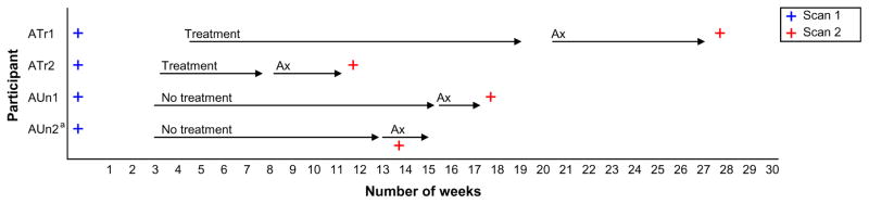
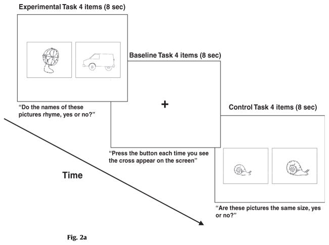
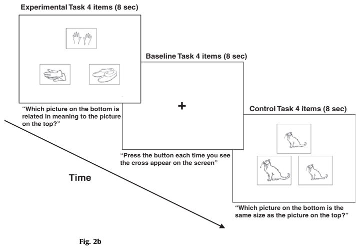
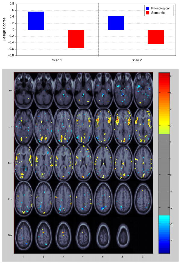
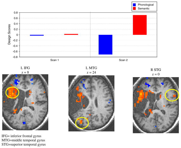
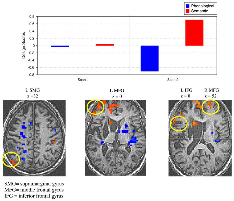
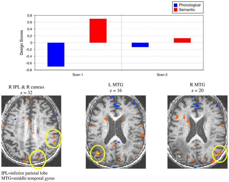
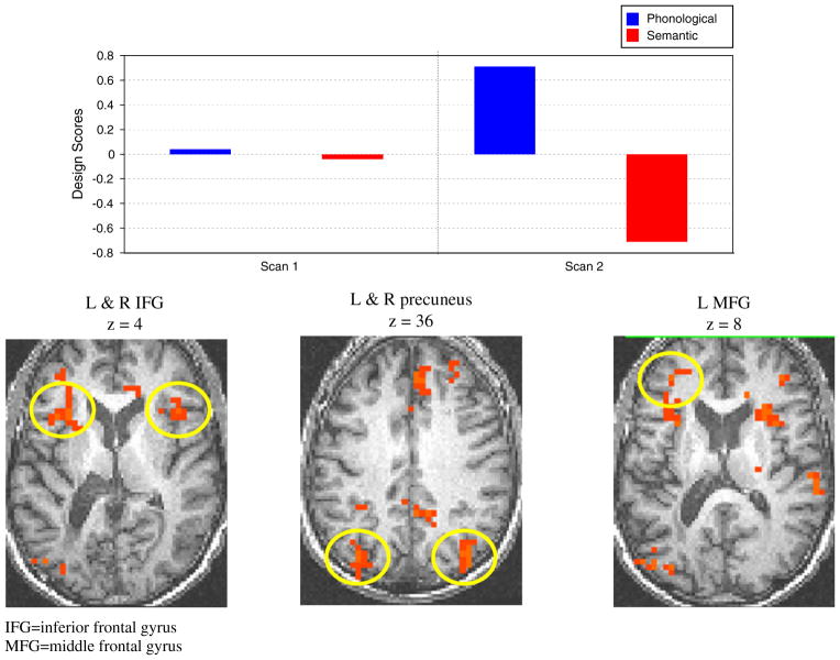
Functional magnetic resonance imaging (fMRI) was used to investigate the neural processing characteristics associated with word retrieval abilities after a phonologically-based treatment for anomia in two stroke patients with aphasia. Neural activity associated with a phonological and a semantic task was compared before and after treatment with fMRI. In addition to the two patients who received treatment, two patients with aphasia who did not receive treatment and 10 healthy controls were also scanned twice. In the two patients who received treatment, both of whose naming improved after treatment, results showed that activation patterns changed after treatment on the semantic task in areas that would have been expected (e.g., left hemisphere frontal and temporal areas). For one control patient, there were no significant changes in brain activation at the second scan; a second control patient showed changes in brain activation at the second scan, on the semantic task, however, these changes were not accompanied with improved performance in naming. In addition, there appeared to be bilateral, or even more right than left hemisphere brain areas activated in this patient than in the treated patients. The healthy control group showed no changes in activation at the second scan. These findings are discussed with reference to the literature on the neural underpinnings of recovery after treatment for anomia in aphasia.
2010 Elsevier Inc. All rights reserved.
Figures
Similar articles
-
Less is more: neural mechanisms underlying anomia treatment in chronic aphasic patients.Brain. 2017 Nov 1;140(11):3039-3054. doi: 10.1093/brain/awx234. Brain. 2017. PMID: 29053773 Free PMC article.
-
Neural underpinnings for model-oriented therapy of aphasic word production.Neuropsychologia. 2014 May;57:154-65. doi: 10.1016/j.neuropsychologia.2014.03.010. Epub 2014 Mar 28. Neuropsychologia. 2014. PMID: 24686092
-
A functional MRI study of the relationship between naming treatment outcomes and resting state functional connectivity in post-stroke aphasia.Hum Brain Mapp. 2014 Aug;35(8):3919-31. doi: 10.1002/hbm.22448. Epub 2014 Jan 22. Hum Brain Mapp. 2014. PMID: 24453137 Free PMC article.
-
Taking Sides: An Integrative Review of the Impact of Laterality and Polarity on Efficacy of Therapeutic Transcranial Direct Current Stimulation for Anomia in Chronic Poststroke Aphasia.Neural Plast. 2016;2016:8428256. doi: 10.1155/2016/8428256. Epub 2015 Dec 27. Neural Plast. 2016. PMID: 26819777 Free PMC article. Review.
-
Anomia training and brain stimulation in chronic aphasia.Neuropsychol Rehabil. 2011 Oct;21(5):717-41. doi: 10.1080/09602011.2011.621275. Neuropsychol Rehabil. 2011. PMID: 22011016 Review.
Cited by
-
Treatment-related changes in neural activation vary according to treatment response and extent of spared tissue in patients with chronic aphasia.Cortex. 2019 Dec;121:147-168. doi: 10.1016/j.cortex.2019.08.016. Epub 2019 Sep 19. Cortex. 2019. PMID: 31627014 Free PMC article.
-
Augmenting melodic intonation therapy with non-invasive brain stimulation to treat impaired left-hemisphere function: two case studies.Front Psychol. 2014 Feb 4;5:37. doi: 10.3389/fpsyg.2014.00037. eCollection 2014. Front Psychol. 2014. PMID: 24550864 Free PMC article.
-
The impact of dose on naming accuracy with persons with aphasia.Aphasiology. 2016;30(9):983-1011. doi: 10.1080/02687038.2015.1100705. Epub 2016 Oct 16. Aphasiology. 2016. PMID: 28133407 Free PMC article.
-
Cognitive Training to Enhance Aphasia Therapy (Co-TrEAT): A Feasibility Study.Front Rehabil Sci. 2022 Apr 5;3:815780. doi: 10.3389/fresc.2022.815780. eCollection 2022. Front Rehabil Sci. 2022. PMID: 36188983 Free PMC article.
-
Evidence of cortical reorganization of language networks after stroke with subacute Broca's aphasia: a blood oxygenation level dependent-functional magnetic resonance imaging study.Neural Regen Res. 2017 Jan;12(1):109-117. doi: 10.4103/1673-5374.198996. Neural Regen Res. 2017. PMID: 28250756 Free PMC article.
References
-
- Baxter LC, Syakin AJ, Flashman LA, Johnson SC, Guerin SJ, Babcock DR, et al. Sex differences in semantic language processing: A functional MRI study. Brain and Language. 2003;84:264–272. - PubMed
-
- Belin P, Van Eeckhout P, Zilbovicius M, Remy P, François C, Guillaum S, et al. Recovery from nonfluent aphasia after melodic intonation therapy: A PET study. Neurology. 1996;47:1504–1511. - PubMed
-
- Breier JI, Maher LM, Schmadeke S, Hasan KM, Papanicolaou AC. Changes in language-specific brain activation after therapy for aphasia using magnetoencephalography: A case study. Neurocase. 2007;13(3):169–177. - PubMed
-
- Busk PL, Serlin R. Meta-analysis for single case research. In: Kratochwill TR, Levin JR, editors. Single-case research design and analysis: New directions for psychology and education. Hillsdale, NJ: Lawrence Erlbaum Associates; 1992.
Publication types
MeSH terms
Grants and funding
LinkOut - more resources
Full Text Sources
