. 2010 Aug 20;285(34):26618-27.
doi: 10.1074/jbc.M109.073155. Epub 2010 Jun 11.

Smad3 regulates Rho signaling via NET1 in the transforming growth factor-beta-induced epithelial-mesenchymal transition of human retinal pigment epithelial cells

Affiliations

Smad3 regulates Rho signaling via NET1 in the transforming growth factor-beta-induced epithelial-mesenchymal transition of human retinal pigment epithelial cells

Jungeun Lee et al. J Biol Chem. .

Abstract

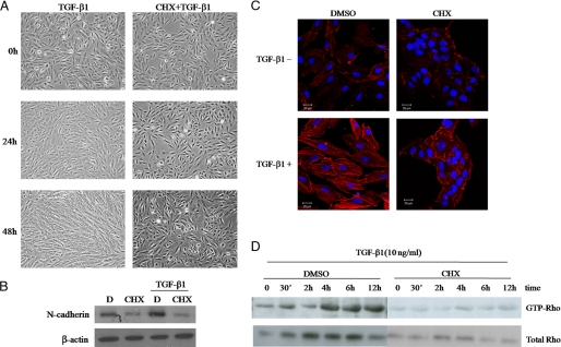
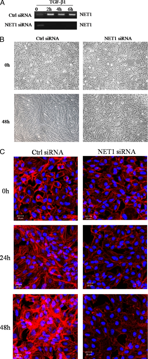
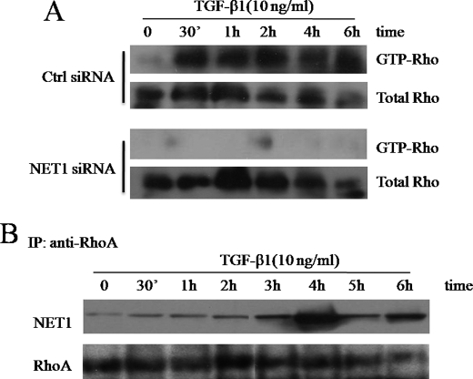
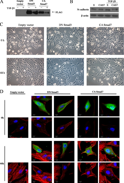
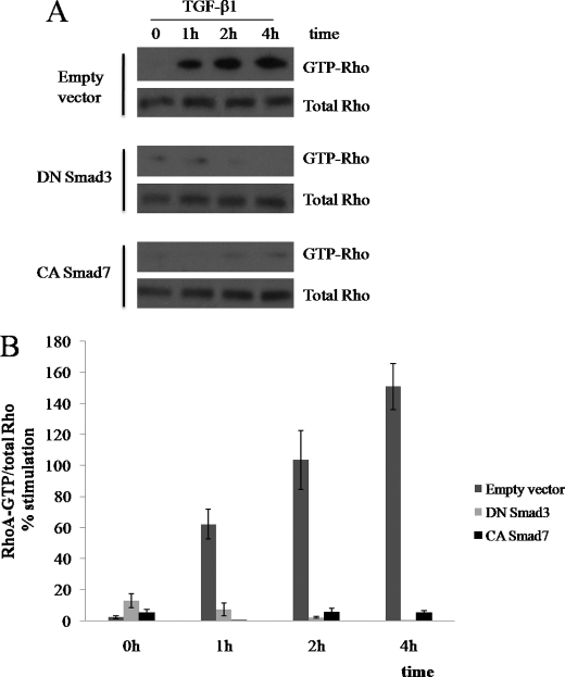
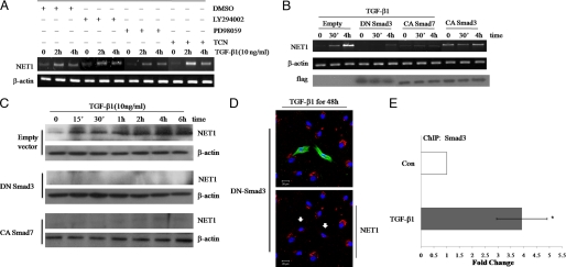
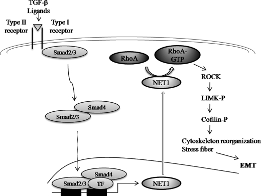
We previously demonstrated that RhoA-dependent signaling regulates transforming growth factor-beta1 (TGF-beta1)-induced cytoskeletal reorganization in the human retinal pigment epithelial cell line ARPE-19. Smad pathways have also been shown to mediate TGF-beta1 activity. Here, we examined what regulates Rho GTPase activity and tested whether Smad signaling cross-talks with Rho pathways during TGF-beta1-induced actin rearrangement. Using small interfering RNAs, we found that NET1, the guanine nucleotide exchange factor of RhoA, is critical for TGF-beta1-induced cytoskeletal reorganization, N-cadherin expression, and RhoA activation. In ARPE-19 cells lacking NET1, TGF-beta1-induced stress fibers and N-cadherin expression were not observed. Interestingly, in dominant-negative Smad3-expressing or constitutively active Smad7 cells, TGF-beta1 failed to induce NET1 mRNA and protein expression. Consistent with these results, both dominant-negative Smad3 and constitutively active Smad7 blocked the cytoplasmic localization of NET1 and inhibited interactions between NET1 and RhoA. Finally, we found that NET1 is a direct gene target of TGF-beta1 via Smad3. Taken together, our results demonstrate that Smad3 regulates RhoA activation and cytoskeletal reorganization by controlling NET1 in TGF-beta1-induced ARPE-19 cells. These data define a new role for Smad3 as a modulator of RhoA activation in the regulation of TGF-beta1-induced epithelial-mesenchymal transitions.


Figures

FIGURE 1.
FIGURE 1.
Protein synthesis is required for TGF-β1-mediated changes in cell morphology and Rho activation. Serum-starved ARPE-19 cells were treated with 10 ng/ml TGF-β1 for 0, 24, or 48 h in the presence or absence of the protein synthesis inhibitor cycloheximide (CHX). D, DMSO. A, the cells were monitored by phase-contrast microscopy at the indicated times. B, shown is a Western blot of N-cadherin under the same treatment conditions as A. C, the actin cytoskeleton was visualized using rhodamine-labeled phalloidin; blue is from Hoechst staining of the nuclei to show all cells. Bar, 20 μm. D, lysed cells were subjected to GST pulldown assays using GST-rhotekin and Western blot analysis using anti-Rho.
FIGURE 2.
FIGURE 2.
TGF-β1 induces NET1 expression in ARPE-19 cells. A, TGF-β1 induces NET1 transcription. Serum-starved ARPE-19 cells were treated with 10 ng/ml TGF-β1 for 0–4 h. After treatment, total RNA was isolated, reverse-transcribed, and amplified by PCR. β-Actin was evaluated as a loading control. B, TGF-β1 induces NET1 protein. Serum-starved ARPE-19 cells were treated with 10 ng/ml TGF-β1 for 0–24 h. Cell lysates were analyzed by Western blotting using a NET1-specific antibody. C, TGF-β1 attenuates cytoplasmic localization of NET1 in ARPE-19 cells. Serum-starved ARPE-19 cells were treated with 10 ng/ml TGF-β1 for 0–6 h. Cells were fixed and stained with anti-NET1 followed by Alexa 488-conjugated rabbit anti-goat and examined by fluorescence microscopy. Bar, 20 μm.
FIGURE 3.
FIGURE 3.
NET1 is critical for TGF-β1-mediated cell morphological changes and stress fiber formation. A, expression of NET1 mRNA was analyzed by RT-PCR at the indicated times. B, NET1 regulates TGF-β1-mediated cell morphological change. ARPE-19 cells were transfected for 12 h with control or NET1-specific siRNA. After transfection, cells were switched to serum-free medium for 3 h and treated with TGF-β1 (10 ng/ml) for 48 h. Cells were monitored by phase-contrast microscopy. C, NET1 knockdown prevents TGF-β1-induced stress fiber formation. ARPE-19 cells were transfected with siRNA and treated with TGF-β1 (10 ng/ml) as described in B. The actin cytoskeleton was visualized by rhodamine-labeled phalloidin, and the blue is from Hoechst staining of the nuclei to show all cells. Bar, 20 μm. Ctrl siRNA, Silencer negative control siRNA.
FIGURE 4.
FIGURE 4.
TGF-β1 induces RhoA activation via NET1. A, NET1 induces RhoA activation. ARPE-19 cells were transfected for 12 h with control or NET1-specific siRNA. After transfection, cells were switched to serum-free medium for 3 h and treated with TGF-β1 (10 ng/ml) for 0–6 h. The cells were then lysed, and the levels of active GTP-Rho in TGF-β1-stimulated ARPE-19 cells were determined by GST pulldown assay using GST-rhotekin and Western blot analysis using an anti-Rho antibody. Ctrl, Silencer negative control siRNA. B, TGF-β1 regulates NET1 binding activity to RhoA. ARPE-19 cells were transfected with siRNA and treated with TGF-β1 (10 ng/ml) as described in A and then lysed. Extracts were immunoprecipitated (IP) with anti-human RhoA, and precipitates were subjected to Western blotting using anti-NET1. As a loading control, aliquots of cell extracts were probed with anti-RhoA.
FIGURE 5.
FIGURE 5.
Smad3 mediates TGF-β1-induced actin rearrangement. FLAG-tagged dominant-negative (DN) Smad3 and FLAG-tagged constitutively active (CA) Smad7 constructs were transfected into ARPE-19 cells. Empty vector was used as a control. After transfection, cells were incubated for 48 h with medium alone or medium containing 10 ng/ml TGF-β1, examined by phase-contrast microscopy (C), and then lysed and subjected to Western blot analysis using anti-FLAG antibodies (A). B, Western blot of N-cadherin under the same treatment conditions as A is shown. E, empty vector; CAS7, constitutively active Smad7. D, ARPE-19 cells were transfected for 12 h with plasmids expressing DN Smad3 or CA Smad7. After transfection, cells were switched to serum-free medium for 3 h and treated with TGF-β1 (10 ng/ml) for 48 h. Cells were fixed and stained with anti-FLAG followed by Alexa 488-conjugated secondary antibody (green) and stained with rhodamine-labeled phalloidin (red). Blue is Hoechst nuclear staining. Pictures were taken under a Zeiss confocal microscope. Bar, 10 μm.
FIGURE 6.
FIGURE 6.
Smad3 mediates TGF-β1-induced RhoA activation. ARPE-19 cells were transfected for 12 h with plasmids expressing dominant-negative (DN) Smad3 or constitutively active (CA) Smad7. Empty vector was used as a control. After transfection, cells were switched to serum-free medium for 3 h and treated with TGF-β1 (10 ng/ml) for 0–4 h. A, cells were then lysed, and active RhoA was precipitated with GST-rhotekin. The total levels of RhoA are also shown. B, statistical analysis of TGF-β1-induced RhoA activation. Error bars represent S.D. *, p < 0.01 compared with black bar within same data set as calculated by Student's t test.
FIGURE 7.
FIGURE 7.
Smad3 regulates RhoA activation through NET1. A, MEK, Akt, and phosphatidylinositol 3-kinase signaling do not affect TGF-β1-induced NET1 mRNA expression. Serum-starved ARPE-19 cells were pretreated for 1 h with vehicle (DMSO) or 10 μm MEK, Akt, or phosphatidylinositol 3-kinase inhibitors (PD98059, Triciribine (TCN), or LY294002, respectively) and then treated with 10 ng/ml TGF-β1 for 0, 2, or 4 h. After treatment, total RNA was isolated, reverse-transcribed, and amplified by PCR. β-Actin was evaluated as a loading control. B, Smad3 regulates NET1 expression. ARPE-19 cells were transfected for 12 h with plasmids expressing dominant-negative (DN) Smad3, constitutively active (CA) Smad3, or constitutively active Smad7. Empty vector was used as a control. After transfection, cells were switched to serum-free medium for 3 h and treated with TGF-β1 (10 ng/ml) for 0, 0.5, or 4 h. After treatment, total RNA was isolated, reverse-transcribed, and amplified by PCR and analyzed by Western blot using a FLAG-specific antibody. C, cells were treated as in B, lysed, and analyzed by Western blot using a NET1-specific antibody. D, Smad3 induces cytoplasmic localization of NET1. ARPE-19 cells were transfected with DNA plasmid and treated with TGF-β1 (10 ng/ml) for 48 h as described in B. Cells were fixed and stained with anti-FLAG followed by Alexa 488 (green) and stained with anti-NET1 followed by Alexa 546 (red). Blue is Hoechst nuclear staining. Pictures were taken under a Zeiss confocal microscope. Arrows indicate transfected cells. Bar, 20 μm. E, NET1 is a Smad3 target gene. ChIP analysis was performed using an anti-Smad3 antibody, and quantitative PCR was performed with primers corresponding to the NET1 promoter region. Cells were left untreated or stimulated with TGF-β1 for 30 min. Quantitative ChIP values are expressed as -fold change in site occupancy and represent the average and S.D. from three independent experiments. Con, control.
FIGURE 8.
FIGURE 8.
Mechanisms of actin cytoskeleton reorganization induced by TGF-β in ARPE-19 cells. Upon stimulation with TGF-β, RhoA can be activated by NET1 through cross-talking with Smad3 to induce actin stress fiber formation during EMT. TF, transcription factor.

References

    1. Bachman K. E., Park B. H. (2005) Curr. Opin. Oncol. 17, 49–54 - PubMed
    1. Lu J., Wu Y., Sousa N., Almeida O. F. (2005) Development 132, 3231–3242 - PubMed
    1. Massagué J. (1998) Annu. Rev. Biochem. 67, 753–791 - PubMed
    1. Song J. (2007) Cell Res. 17, 289–290 - PubMed
    1. Yang Y., Pan X., Lei W., Wang J., Song J. (2006) Oncogene 25, 7235–7244 - PubMed

Publication types

MeSH terms