. 2010 Aug 20;285(34):25928-40.
doi: 10.1074/jbc.M110.117911. Epub 2010 Jun 11.

Cross-talk between the p38alpha and JNK MAPK pathways mediated by MAP kinase phosphatase-1 determines cellular sensitivity to UV radiation

Affiliations

Cross-talk between the p38alpha and JNK MAPK pathways mediated by MAP kinase phosphatase-1 determines cellular sensitivity to UV radiation

Christopher J Staples et al. J Biol Chem. .

Erratum in

  • J Biol Chem. 2011 May 13;286(19):17398

Abstract

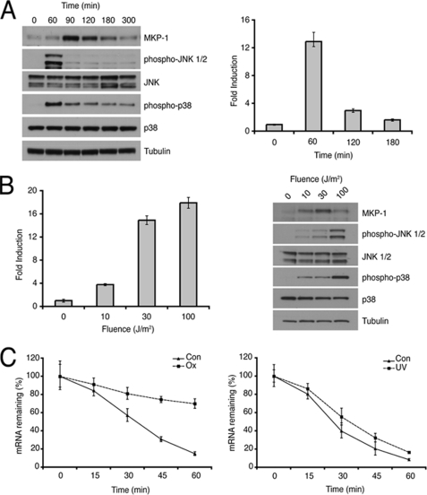
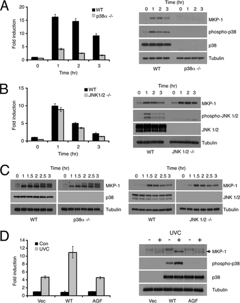
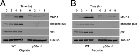
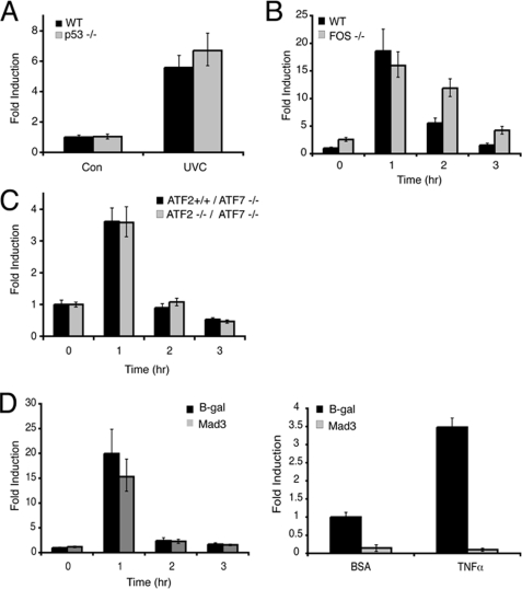
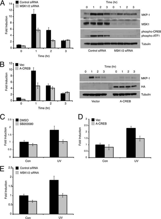
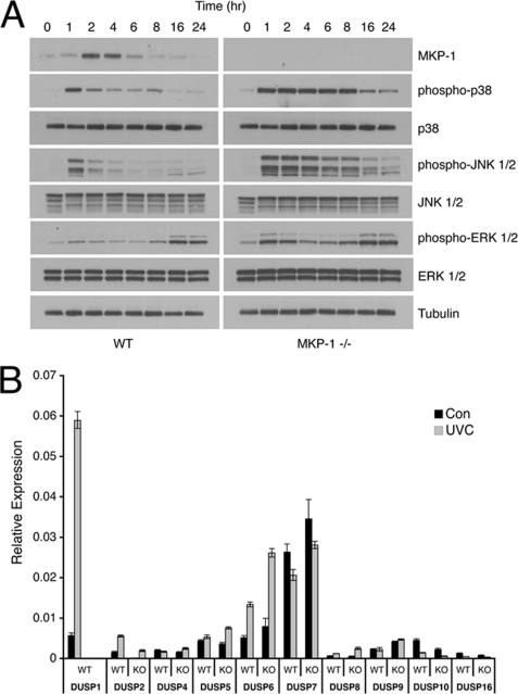
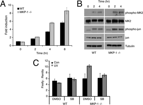
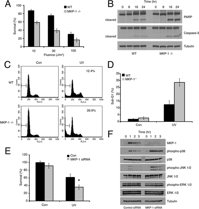
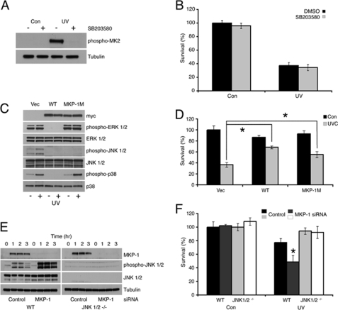
MAPK phosphatase-1 (DUSP1/MKP-1) is a mitogen and stress-inducible dual specificity protein phosphatase, which can inactivate all three major classes of MAPK in mammalian cells. DUSP1/MKP-1 is implicated in cellular protection against a variety of genotoxic insults including hydrogen peroxide, ionizing radiation, and cisplatin, but its role in the interplay between different MAPK pathways in determining cell death and survival is not fully understood. We have used pharmacological and genetic tools to demonstrate that DUSP1/MKP-1 is an essential non-redundant regulator of UV-induced cell death in mouse embryo fibroblasts (MEFs). The induction of DUSP1/MKP-1 mRNA and protein in response to UV radiation is mediated by activation of the p38alpha but not the JNK1 or JNK2 MAPK pathways. Furthermore, we identify MSK1 and -2 and their downstream effectors cAMP-response element-binding protein/ATF1 as mediators of UV-induced p38alpha-dependent DUSP1/MKP-1 transcription. Dusp1/Mkp-1 null MEFs display increased signaling through both the p38alpha and JNK MAPK pathways and are acutely sensitive to UV-induced apoptosis. This lethality is rescued by the reintroduction of wild-type DUSP1/MKP-1 and by a mutant of DUSP1/MKP-1, which is unable to bind to either p38alpha or ERK1/2, but retains full activity toward JNK. Importantly, whereas small interfering RNA-mediated knockdown of DUSP1/MKP-1 sensitizes wild-type MEFs to UV radiation, DUSP1/MKP-1 knockdown in MEFS lacking JNK1 and -2 does not result in increased cell death. Our results demonstrate that cross-talk between the p38alpha and JNK pathways mediated by induction of DUSP1/MKP-1 regulates the cellular response to UV radiation.


Figures

FIGURE 1.
FIGURE 1.
Induction of DUSP1/MKP-1 by UV radiation. A, wild-type MEFs were either mock irradiated or exposed to 30 J/m2 UV and then incubated at 37 °C for the indicated times. Cells were then lysed, and proteins analyzed by SDS-PAGE and Western blotting using the indicated antibodies (left). DUSP1/MKP-1 mRNA levels were also determined using quantitative RT-PCR (right). B, wild-type MEFs were exposed to 0, 10, 30, or 100 J/m2 UV and incubated for 1 h at 37 °C. Cells were then lysed, and DUSP1/MKP-1 mRNA levels were determined using quantitative RT-PCR (left). Proteins were also analyzed by SDS-PAGE and Western blotting using the indicated antibodies (right). C, wild-type MEFs were either treated with 300 μm H2O2 (Ox) for 3 h or exposed to 30 J/m2 UV (UV) and incubated for 1 h, before addition of 2 μg/ml of actinomycin D. Cells were then lysed at the indicated times, and DUSP1/MKP-1 mRNA levels were determined using quantitative RT-PCR. Experiments were performed three times, each with triplicate determinations, and mean values with associated errors are shown (mean ± S.E.).
FIGURE 2.
FIGURE 2.
DUSP1/MKP-1 induction by UV is mediated by p38α. A, wild-type (WT) or p38α−/− MEFs were either mock irradiated or exposed to 30 J/m2 UV and then incubated at 37 °C for the indicated times. Cells were then lysed, and DUSP1/MKP-1 mRNA levels were determined using quantitative RT-PCR (left). Proteins were also analyzed by SDS-PAGE and Western blotting using the indicated antibodies (right). B, wild-type (WT) or Jnk1/2−/− MEFs were either mock irradiated or exposed to 30 J/m2 UV and then incubated at 37 °C for the indicated times. Cells were then lysed, and DUSP1/MKP-1 mRNA levels were determined using quantitative RT-PCR (left). Proteins were also analyzed by SDS-PAGE and Western blotting using the indicated antibodies (right). C, wild-type, p38α−/−, or Jnk1/2−/− MEFs were starved for 16 h in medium containing 0.5% FBS, then stimulated with 15% FBS for the indicated times. Cells were then lysed, and proteins analyzed by SDS-PAGE and Western blotting using the indicated antibodies. D, p38α−/− MEFs were transfected with either empty vector, or expression vectors (Vec) encoding either wild-type p38α or a non-activable (AGF) mutant of p38α. Cells were mock irradiated or exposed to 30 J/m2 UV and then incubated at 37 °C for 1 h before lysis. DUSP1/MKP-1 mRNA levels were determined using quantitative RT-PCR (left). Proteins were also analyzed by SDS-PAGE and Western blotting using the indicated antibodies (right). Experiments were performed three times, each in triplicate, and mean values with associated errors are shown (mean ± S.E.).
FIGURE 3.
FIGURE 3.
DUSP1/MKP-1 induction by both cisplatin and oxidative stress is mediated by p38α. A, wild-type (WT) or p38α−/− MEFs were treated with 100 μm cisplatin for the indicated times. B, wild-type or p38α−/− MEFs were treated with 300 μm hydrogen peroxide for the indicated times. Cells were then lysed, and proteins analyzed by SDS-PAGE and Western blotting using the indicated antibodies. Data are representative of three independent experiments.
FIGURE 4.
FIGURE 4.
DUSP1/MKP-1 induction by UV is independent of p53, c-Fos, ATF2, and NF-κB. A, HCT116 p53+/+, and p53−/− cells were either mock irradiated or exposed to 100 J/m2 UV and then incubated at 37 °C for 1 h. B, wild-type (WT) and c-Fos−/− MEFs were either mock irradiated or exposed to 30 J/m2 UV and then incubated at 37 °C for the indicated times. C, Atf7−/−/Atf2+/+ and Atf7−/−/Atf2−/− MEFs were either mock irradiated or exposed to 30 J/m2 UV and then incubated at 37 °C for the indicated times. D, wild-type MEFs were transfected with either empty vector, or an expression vector encoding Mad3. After 24 h, cells were exposed to 30 J/m2 UV and then incubated at 37 °C for the indicated times. DUSP1/MKP-1 mRNA levels were assessed by quantitative RT-PCR (left). Wild-type MEFs were cotransfected with the NFB reporter construct 3xκB-ConA-luc and either an empty vector or an expression vector encoding Mad3. Renilla luciferase was included as a transfection control. Cells were then stimulated with either 10 ng/ml of bovine serum albumin (BSA) or tumor necrosis factor (TNF) α. After 8 h, cells were lysed, and luciferase activities were determined (right). Experiments were performed three times, each with triplicate determinations and mean values with associated errors are shown (mean ± S.E.). B-gal, β-galactosidase.
FIGURE 5.
FIGURE 5.
Induction of DUSP1/MKP-1 by UV is dependent on signaling via MSK1/2 and CREB/ATF1. A, wild-type MEFs were transfected with either a control siRNA, or siRNAs targeting MSK1 and MSK2. Cells were either mock irradiated or exposed to 30 J/m2 UV and then incubated at 37 °C for the indicated times. Cells were then lysed and either DUSP1/MKP-1 mRNA levels were determined using quantitative RT-PCR (left) or proteins were analyzed by SDS-PAGE and Western blotting using the indicated antibodies (right). B, wild-type MEFs were transfected with either empty vector or an expression vector (Vec) encoding hemagglutinin-tagged A-CREB. After 24 h, cells were either mock irradiated or exposed to 30 J/m2 UV and then incubated at 37 °C for the indicated times. Cells were then lysed and either DUSP1/MKP-1 mRNA levels were determined using quantitative RT-PCR (left) or proteins were analyzed by SDS-PAGE and Western blotting using the indicated antibodies (right). Wild-type MEFs stably transfected with the MKP-1 reporter pGL4.28-MKP-1-luc were either pre-treated for 1 h with dimethyl sulfoxide (DMSO) or 10 μm SB203580 (C), transfected with either empty vector or an expression vector encoding A-CREB (D), or transfected with either a control siRNA or siRNAs targeting MSK1 and -2 (E). Cells were then either mock irradiated or exposed to 30 J/m2 UV and incubated at 37 °C for 8 h. Cells were then lysed, firefly luciferase activity was quantified and its levels normalized to protein concentration. Experiments were performed three times, each with triplicate determinations, and mean values with associated errors are shown (mean ± S.E.). DMSO, dimethyl sulfoxide; HA, hemagglutinin.
FIGURE 6.
FIGURE 6.
DUSP1/MKP-1 is an essential non-redundant regulator of UV-induced p38α and JNK signaling. A, wild-type (WT) or Dusp1/Mkp-1−/− MEFs were either mock irradiated or exposed to 30 J/m2 UV and then incubated at 37 °C for the indicated times. Cells were then lysed, and proteins analyzed by SDS-PAGE and Western blotting using the indicated antibodies. B, WT or Dusp1/Mkp-1−/− MEFs were either mock irradiated or exposed to 30 J/m2 UV and then incubated at 37 °C for 1 h. DUSP/MKP mRNA levels were determined using quantitative RT-PCR. Experiments were performed three times, each with triplicate determinations, and mean values with associated errors are shown (mean ± S.E.). KO, knock-out.
FIGURE 7.
FIGURE 7.
Loss of DUSP1/MKP-1 results in increased MAPK signaling and gene transcription. A, wild-type (WT) or Dusp1/Mkp-1−/− MEFs were either mock irradiated or exposed to 30 J/m2 UV and then incubated at 37 °C for the indicated times. c-Fos mRNA levels were determined using quantitative RT-PCR. B, wild-type or Dusp1/Mkp-1−/− MEFs were either mock irradiated or exposed to 30 J/m2 UV and then incubated at 37 °C for the indicated times. Cells were lysed, and proteins analyzed by SDS-PAGE followed by Western blotting using the indicated antibodies. C, wild-type or Dusp1/Mkp-1−/− MEFs were cotransfected with a firefly luciferase reporter driven by the proximal 3-kb region of the DUSP1/MKP-1 promoter and as a transfection control a plasmid encoding Renilla luciferase. After 24 h, cells were preincubated with either dimethyl sulfoxide (DMSO) or 10 μm SB203580 (SB), exposed to 30 J/m2 UV, and then incubated at 37 °C for 5 h. Cells were lysed, and luciferase activities were determined. Experiments were performed three times, each with triplicate determinations and mean values with associated errors are shown (mean ± S.E.).
FIGURE 8.
FIGURE 8.
Loss of DUSP1/MKP-1 sensitizes cells to UV-induced apoptosis. A, wild-type (WT) or Dusp1/Mkp-1−/− MEFs were exposed to 0, 10, 30, or 100 J/m2 UV, and then incubated at 37 °C for 36 h. Cell survival was assessed by MTS assay, and results were normalized to untreated control values. B, wild-type or Dusp1/Mkp-1−/− MEFs were exposed to 30 J/m2 UV, and then incubated at 37 °C for the indicated times. Cells were lysed, and proteins analyzed by SDS-PAGE followed by Western blotting using the indicated antibodies. C and D, wild-type or Dusp1/Mkp-1−/− MEFs were exposed to 30 J/m2 UV, and then incubated at 37 °C for 48 h. Cells were fixed in ethanol and stained using propidium iodide. Cell death was assessed by flow cytometry. E, wild-type MEFs were transfected with either a control siRNA, or an siRNA targeting DUSP1/MKP-1. After 48 h, cells were exposed to 30 J/m2 UV. Cell survival was assessed after 36 h by MTS assay, and results were normalized to untreated control values. F, wild-type MEFs were transfected as in E, exposed to 30 J/m2 UV, and then incubated at 37 °C for the indicated times. Cells were then lysed, and proteins analyzed by SDS-PAGE followed by Western blotting using the indicated antibodies. Experiments were performed three times, with triplicate determinations, and mean values with associated errors are shown (mean ± S.E.). * = p < 0.05, comparing the survival of UV-irradiated cells transfected with either control or DUSP1/MKP-1 siRNA using the Student's t test. PARP, poly(ADP-ribose) polymerase.
FIGURE 9.
FIGURE 9.
UV-induced cell death is JNK dependent. A, Dusp1/Mkp-1−/− MEFs were pre-treated with dimethyl sulfoxide (DMSO) or 10 μm SB203580, and then exposed to 30 J/m2 UV. Following incubation at 37 °C for 1 h, cells were lysed, and proteins analyzed by SDS-PAGE followed by Western blotting using the indicated antibodies. B, Dusp1/Mkp-1−/− MEFs were treated as in A. After 36 h, cell survival was assessed by MTS assay and the results normalized to untreated control values. C, Dusp1/Mkp-1−/− MEFs were transfected with either empty vector or an expression vector encoding either wild-type (WT) MKP-1 or MKP-1 M. After 24 h, cells were exposed to 30 J/m2 UV and incubated at 37 °C for 1 h. Cells were then lysed, and proteins analyzed by SDS-PAGE followed by Western blotting using the indicated antibodies. D, Dusp1/Mkp-1−/− MEFs were transfected as in C. After 24 h, cells were exposed to 30 J/m2 UV. Following incubation at 37 °C for 36 h, cell survival was assessed by MTS assay, and the results were normalized to untreated control values. * = p < 0.05, comparing the survival of UV-irradiated cells transfected with empty vector with those transfected with either an expression vector encoding WT MKP-1, or those transfected with an expression vector encoding MKP-1 M using Student's t test. E, wild-type and Jnk1/2−/− MEFs were transfected with either a control siRNA, or an siRNA targeting DUSP1/MKP-1. After 48 h, cells were either mock irradiated or exposed to 30 J/m2 UV, and then incubated at 37 °C for the indicated times. Cells were lysed, and proteins analyzed by SDS-PAGE followed by Western blotting using the indicated antibodies. F, wild-type and Jnk1/2−/− MEFs were transfected as in E, then either mock irradiated or exposed to 30 J/m2 UV. Cell survival was assessed after 36 h by MTS assay, and results were normalized to untreated control values. * = p < 0.05, comparing the survival of UV-irradiated cells transfected with control siRNA and those transfected with an siRNA targeting MKP-1 using Student's t test. Experiments were performed three times, each with triplicate determinations, and mean values with associated errors are shown (mean ± S.E.).

Similar articles

Cited by

References

    1. Dickinson R. J., Keyse S. M. (2006) J. Cell Sci. 119, 4607–4615 - PubMed
    1. Owens D. M., Keyse S. M. (2007) Oncogene 26, 3203–3213 - PubMed
    1. Chu Y., Solski P. A., Khosravi-Far R., Der C. J., Kelly K. (1996) J. Biol. Chem. 271, 6497–6501 - PubMed
    1. Groom L. A., Sneddon A. A., Alessi D. R., Dowd S., Keyse S. M. (1996) EMBO J. 15, 3621–3632 - PMC - PubMed
    1. Slack D. N., Seternes O. M., Gabrielsen M., Keyse S. M. (2001) J. Biol. Chem. 276, 16491–16500 - PubMed

Publication types

MeSH terms

Substances