Randomized Controlled Trial
. 2010 Jul;126(1):53-61.
doi: 10.1542/peds.2009-2800. Epub 2010 Jun 14.

Incidental findings on brain magnetic resonance imaging of children with sickle cell disease

Collaborators, Affiliations
Randomized Controlled Trial

Incidental findings on brain magnetic resonance imaging of children with sickle cell disease

Lori C Jordan et al. Pediatrics. 2010 Jul.

Abstract

Objective: We describe the prevalence and range of incidental intracranial abnormalities identified through MRI of the brain in a large group of children screened for a clinical trial.

Methods: We included 953 children between 5 and 14 years of age who were screened with MRI of the brain for the Silent Infarct Transfusion Trial. All had sickle cell anemia or sickle beta-null thalassemia. MRI scans were interpreted by 3 neuroradiologists. MRI scans reported to have any abnormality were reviewed by 2 study neuroradiologists. Incidental findings were classified into 4 categories, that is, no, routine, urgent, or immediate referral recommended. Cerebral infarctions and vascular lesions were not considered incidental and were excluded.

Results: We identified 63 children (6.6% [95% confidence interval: 5.1%-8.4%]) with 68 incidental intracranial MRI findings. Findings were classified as urgent in 6 cases (0.6%), routine in 25 cases (2.6%), and no referral required in 32 cases (3.4%). No children required immediate referral. Two children with urgent findings underwent surgery in the subsequent 6 months.

Conclusion: In this large cohort of children, incidental intracranial findings were identified for 6.6%, with potentially serious or urgent findings for 0.6%.


Figures
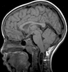
Figure 1(a-f)
Figure 1(a-f)
Urgent Incidental MRI Findings. 1a) Sagittal T1 view of an incidental Chiari I malformation (arrow head) with cervical spinal cord syrinx (arrow). 1b) Sagittal T1 view of an incidental Chiari I malformation (arrow head) with cervical spinal cord syrinx containing plicae or septations (arrow). 1c) Axial FLAIR image with an incidentally detected cystic lesion in the right temporal lobe that could represent a tumor. Arrows point to the cyst and an area of nodularity within the cyst. 1d) T1 sagittal and T2 axial views of an incidental tectal lesion, possible glioma (arrows). 1e) Axial T2 and coronal FLAIR images of an incidental corpus callosum lesion, potentially representing a tumor or an infarct (arrows). 1f) Axial FLAIR image of a left hippocampal cystic lesion (arrow), possible tumor versus a focus of dysplasia.
Figure 1(a-f)
Figure 1(a-f)
Urgent Incidental MRI Findings. 1a) Sagittal T1 view of an incidental Chiari I malformation (arrow head) with cervical spinal cord syrinx (arrow). 1b) Sagittal T1 view of an incidental Chiari I malformation (arrow head) with cervical spinal cord syrinx containing plicae or septations (arrow). 1c) Axial FLAIR image with an incidentally detected cystic lesion in the right temporal lobe that could represent a tumor. Arrows point to the cyst and an area of nodularity within the cyst. 1d) T1 sagittal and T2 axial views of an incidental tectal lesion, possible glioma (arrows). 1e) Axial T2 and coronal FLAIR images of an incidental corpus callosum lesion, potentially representing a tumor or an infarct (arrows). 1f) Axial FLAIR image of a left hippocampal cystic lesion (arrow), possible tumor versus a focus of dysplasia.
Figure 2
Figure 2
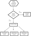
Flow Chart of Incidental Findings in Children Screened for the SIT Trial.

References

    1. Katzman GL, Dagher AP, Patronas NJ. Incidental findings on brain magnetic resonance imaging from 1000 asymptomatic volunteers. JAMA. 1999;282(1):36–39. - PubMed
    1. Weber F, Knopf H. Incidental findings in magnetic resonance imaging of the brains of healthy young men. J Neurol Sci. 2006;240(1-2):81–84. - PubMed
    1. Yue NC, Longstreth WT, Jr, Elster AD, Jungreis CA, O’Leary DH, Poirier VC. Clinically serious abnormalities found incidentally at MR imaging of the brain: Data from the cardiovascular health study. Radiology. 1997;202(1):41–46. - PubMed
    1. Vernooij MW, Ikram MA, Tanghe HL, et al. Incidental findings on brain MRI in the general population. N Engl J Med. 2007;357(18):1821–1828. - PubMed
    1. Morris Z, Whiteley WN, Longstreth WT, Jr, et al. Incidental findings on brain magnetic resonance imaging: systematic review and meta-analysis. BMJ. 2009;339:b3016. - PMC - PubMed

Publication types

MeSH terms