. 2010 Aug 15;588(Pt 16):3031-43.
doi: 10.1113/jphysiol.2009.184705. Epub 2010 Jun 14.

Subthreshold membrane potential oscillations in inferior olive neurons are dynamically regulated by P/Q- and T-type calcium channels: a study in mutant mice

Affiliations

Subthreshold membrane potential oscillations in inferior olive neurons are dynamically regulated by P/Q- and T-type calcium channels: a study in mutant mice

Soonwook Choi et al. J Physiol. .

Abstract

The role of P/Q- and T-type calcium channels in the rhythmic oscillatory behaviour of inferior olive (IO) neurons was investigated in mutant mice. Mice lacking either the CaV2.1 gene of the pore-forming alpha1A subunit for P/Q-type calcium channel, or the CaV3.1 gene of the pore-forming alpha1G subunit for T-type calcium channel were used. In vitro intracellular recording from IO neurons reveals that the amplitude and frequency of sinusoidal subthreshold oscillations (SSTOs) were reduced in the CaV2.1-/- mice. In the CaV3.1-/- mice, IO neurons also showed altered patterns of SSTOs and the probability of SSTO generation was significantly lower (15%, 5 of 34 neurons) than that of wild-type (78%, 31 of 40 neurons) or CaV2.1-/- mice (73%, 22 of 30 neurons). In addition, the low-threshold calcium spike and the sustained endogenous oscillation following rebound potentials were absent in IO neurons from CaV3.1-/- mice. Moreover, the phase-reset dynamics of oscillatory properties of single neurons and neuronal clusters in IO were remarkably altered in both CaV2.1-/- and CaV3.1-/- mice. These results suggest that both alpha1A P/Q- and alpha1G T-type calcium channels are required for the dynamic control of neuronal oscillations in the IO. These findings were supported by results from a mathematical IO neuronal model that incorporated T and P/Q channel kinetics.


Figures
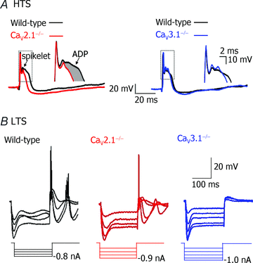
Figure 1
Figure 1. Responses of inferior (IO) neurons to depolarizing or hyperpolarizing current injection in wild-type (black) mice lacking CaV2.1 (red), and mice lacking CaV3.1 (blue)
A, the suprathreshold depolarizing pulse elicited a fast sodium spike followed by a high-threshold calcium spike and an afterhyperpolarization in brainstem slices from a wild-type (black), CaV2.1−/− (red) or, CaV3.1−/− (blue) mouse. Note that the duration and peak of the high-threshold spike was decreased in IO neuron from the CaV2.1−/− mouse (red). The insets on the right of the spikes show details of the afterdepolarizatio duration and spikelet character of the wild-type vs. CaV2.1−/− and CaV3.1−/−. B, responses to a set of hyperpolarizing current pulses in brainstem slice from a wild-type (black), CaV2.1−/− (red) or CaV3.1−/− (blue) mouse. Note that a low-threshold calcium spike was absent in the neuron from the CaV3.1−/− mouse while the high-threshold spike was unaffected. The hyperpolarization-activated cation current was present in slices from all the animals.
Figure 2
Figure 2. Sinusoidal subthreshold oscillations (SSTOs) and rebound potentials in wild-type, CaV2.1−/− and CaV3.1−/− mice
A, representative SSTOs at five membrane potentials (at left of each trace) in wild-type, CaV2.1−/− and CaV3.1−/− mice in the presence of TTX. Oscillations were present at all membrane potential levels in all genotypes, although they were lowest in CaV3.1−/− mice. B, SSTO amplitude plotted as a function of cell membrane potential. (Mean amplitudes were scaled to the largest response taken as 100%.) Note that SSTO amplitude is modulated in wild-type and CaV2.1−/−, but not CaV3.1−/− mice. C, SSTO frequency as a function of cell membrane potential. Note also that frequency was lower in the mutant mice, and that frequency was insensitive to membrane potential in wild-type and mutant mice. Data in B, C and D were obtained from the same cells. D, the intracellular injection of a hyperpolarizing current pulse (−0.8 nA, 0.2–0.3 s) from the resting or hyperpolarized membrane potentials elicited a low-threshold spike and rhythmic oscillations in IO neurons from wild-type (black) and CaV2.1−/− (red), but not CaV3.1−/− (blue) mice, although the rebound activity mediated by the hyperpolarization-activated cation current (Ih) was present.
Figure 3
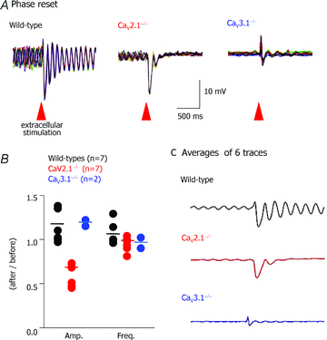
Figure 3. Extracellular stimulation-induced phase reset of SSTOs in single IO neurons from WT, CaV2.1−/− and CaV3.1−/− mice
A, compared to phase reset in wild-type mice, this phenomenon was reduced in CaV3.1−/− and absent in CaV2.1−/− mice (superposition of 6 traces, stimulation 0.1 to ∼2 mA, 0.2 ms). B, plot of ratio of mean amplitude or frequency after/before stimulation in IO cells from wild-type and mutant mice. Only the amplitude of SSTOs in CaV2.1−/− mice was significantly reduced after extracellular stimulation (P < 0.05 by Student's t test). C, mean SSTOs in wild-type (n = 7) and mutant mice showing phase reset in wild-type and CaV3.1−/− (n = 2), but not CaV2.1−/− mice.
Figure 4
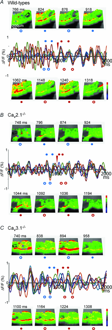
Figure 4. Synchronized oscillations of clusters of IO neurons in wild-type and mutant mice
A, middle row, oscillations before and after stimulus was delivered (filament in first image and at red arrowhead in traces). Blue marks correspond to time images taken before stimulation, red marks to images taken after stimulation at the oscillation troughs (open circles) or peaks (filled circles). Top row, images of IO region of brainstem slice before stimulation. Bottom row, images taken after stimulation. Note increased coherence of cluster activity at peaks compared to troughs, and after compared to before, stimulation (in WT mice, n = 11). B, same as in A for CaV2.1−/− mutant (n = 4). Note near absence of clusters of activity before and modest increase after stimulation. C, same as in A for CaV3.1−/− mutant (n = 6). Note near absence of clusters of activity before and modest increase after stimulation. Three-dimensional images at each time point were superimposed on a contrast photo (100 × 100 pixels, field of view: 5 × 5 mm) of the slice. Voltage changes were recorded from the entire IO. Colour intensity code: 0 (blue) to 255 (red). Reverse FFT analysis was performed from the recordings of oscillation at six points (3 × 3 round pixel) of each slice and shown as coloured traces.
Figure 5
Figure 5. Characteristic estimates of power density for subthreshold oscillations in neurons with P/Q and T channels (wild-type), with only T channels (Cav2.1−/−) or with only PQ channels (Cav3.1−/−)
Red and blue lines in A–C refer to membrane potential depolarization and hyperpolarization. A, wild-type neurons in case of depolarization and hyperpolarization. B, Cav2.1−/− neurons were less sensitive to transition from depolarization to hyperpolarization than wild-type neurons. C, CaV3.1−/− neurons were less sensitive to transition from depolarization to hyperpolarization than wild-type or CaV2.1−/− neurons and the oscillations were also less capable of preserving the SSTO frequency than in both other types of neurons. CaV3.1−/− neurons had less than ∼30% of the activated channels than in wild-type cells, while with T-type channels only the drop is down to ∼70% of the original. D, a qualitative estimate of the probability that channelsof different types are activated depending on the state (depolarization and hyperpolarization states are mirrored for different channels). PQ-type channels were found to operate in much narrow range than T-type channels and therefore their S curve cumulative distribution is much closer to a Step function. Area under appropriate frequency peaks reflects amount of energy it carries.
Figure 6
Figure 6. Effect of added channel ‘noise’ on membrane potential oscillations in wild-type model neurons
Vertical axis is voltage, horizontal axis is time: both values are in conditional model units. A, oscillations in the presence of a low level of noise. B, oscillations when the added noise level is optimal, causing stochastic resonance. C, oscillations in the presence of a high level of channel noise. Note that the regularity of the oscillations is very sensitive to the level of channel noise in the model.

Similar articles

Cited by

References

    1. Bal T, McCormick DA. Synchronized oscillations in the inferior olive are controlled by the hyperpolarization-activated cation current Ih. J Neurophysiol. 1997;77:3145–3156. - PubMed
    1. Bell C, Kawasaki T. Relations among climbing fiber responses of nearby Purkinje cells. J Neurophysiol. 1972;35:155–169. - PubMed
    1. Benardo L, Foster R. Oscillatory behavior in inferior olive neurons: mechanism, modulation, cell aggregates. Brain Res Bull. 1986;17:773–784. - PubMed
    1. Bezrukov S, Vodyanoy I. Stochastic resonance and small-amplitude signal transduction in voltage-gated ion channels. In: Walleczek J, editor. Self-organized Biological Dynamics and Nonlinear Control. Cambridge: Cambridge University Press; 2000.
    1. Bulsara A, Hanggi P, Marchesoni F, Moss F, Shlesinger M. Special issue: Stochastic resonance in physics and biology. J Stat Phys. 1993;70:1–512.

Publication types

MeSH terms

Substances

LinkOut - more resources