. 2010 Sep;78(9):3660-8.
doi: 10.1128/IAI.00386-10. Epub 2010 Jun 14.

Frameshift mutations in a single novel virulence factor alter the in vivo pathogenicity of Chlamydia trachomatis for the female murine genital tract

Affiliations

Frameshift mutations in a single novel virulence factor alter the in vivo pathogenicity of Chlamydia trachomatis for the female murine genital tract

Gail L Sturdevant et al. Infect Immun. 2010 Sep.

Abstract

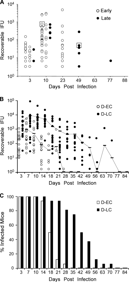
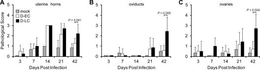
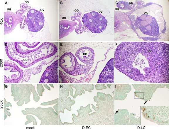
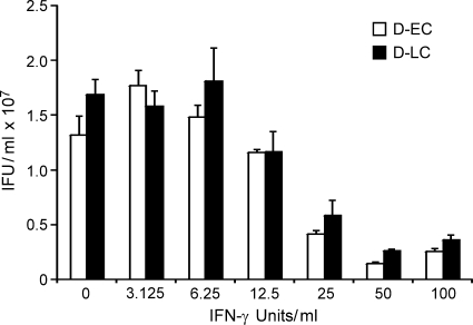
Chlamydia trachomatis is a human pathogen of global importance. An obstacle to studying the pathophysiology of human chlamydial disease is the lack of a suitable murine model of C. trachomatis infection. Mice are less susceptible to infection with human isolates due in part to innate mouse-specific host defense mechanisms to which human strains are sensitive. Another possible factor that influences the susceptibility of mice to infection is that human isolates are commonly cultivated in vitro prior to infection of mice; therefore, virulence genes could be lost as a consequence of negative selective pressure. We tested this hypothesis by infecting innate immunity-deficient C3H/HeJ female mice intravaginally with a human serovar D urogenital isolate that had undergone multiple in vitro passages. We observed early and late infection clearance phenotypes. Strains of each phenotype were isolated and then used to reinfect naïve mice. Following infection, the late-clearance strain was significantly more virulent. It caused unvarying infections of much longer durations with greater infectious burdens that naturally ascended to the upper genital tract, causing salpingitis. Despite contrasting in vivo virulence characteristics, the strains exhibited no differences in the results of in vitro infectivity assays or sensitivities to gamma interferon. Genome sequencing of the strains revealed mutations that localized to a single gene (CT135), implicating it as a critical virulence factor. Mutations in CT135 were not unique to serovar D but were also found in multiple oculogenital reference strains. Our findings provide new information about the pathogenomics of chlamydial infection and insights for improving murine models of infection using human strains.


Figures

FIG. 1.
FIG. 1.
In vivo isolation of C. trachomatis infection phenotypes. (A) Fifty-six C3H/HeJ mice were infected with 105 IFU parental strain D/UW-3/CX, and the course of infection was monitored by culture. The results for culture-positive mice are plotted. Infection durations varied from early (10 to 23 days; open circles) to late (49 to 77 days; closed circles). Chlamydiae were isolated from a single early-clearance (D-EC) or late-clearance (D-LC) strain-infected mouse (boxed) and serially passed in McCoy cells (five passages) to obtain sufficient amounts of organisms to rechallenge naïve animals. (B) Sixteen mice were infected with 105 IFU of D-EC or D-LC, and the course of the infection was monitored by culture. Each data point indicates the number of IFU cultured from a single mouse infected with either D-EC (open circle) or D-LC (filled circle). The results for culture-positive mice are plotted. Samples from mice were cultured until inclusion-negative samples were obtained at two successive time points. Contiguous lines indicate the 16-mouse mean IFU values for both groups and include the results for both positive and negative cultures. Mice infected with the individual isolates exhibited distinctly separate infection and clearance kinetics, suggesting that the in vivo-selected strains were clonal. (C) Data for the same groups whose results are shown in panel A plotted as the percentage of infected mice. The percentage of infected mice in the D-LC group was consistently greater in the later culture periods (days 18 to 42).
FIG. 2.
FIG. 2.
Inflammatory response in the upper genital tract of C. trachomatis-infected mice. Seventy-three C3H/HeJ mice were infected intravaginally with 105 IFU of D-EC, D-LC, or SPG buffer (mock infection). At five different times following infection, the genital tracts were excised and placed in 10% formalin, embedded into paraffin, sliced, and H&E stained. Upper genital tract tissues were examined for chronic inflammatory cellular infiltrates and scored accordingly: 0 for none, 1 for minimal, 2 for mild, 3 for moderate, and 4 for severe. Scoring was performed for the uterine horn (A), oviduct (B), and ovaries (C). The numbers of mock-, D-EC-, and D-LC-infected mice scored ranged from three to seven mice per group. Mean pathological scores are plotted, and error bars indicate the standard error of the mean. Tukey-Kramer pairwise statistical tests were performed at each time point. P values of less than 0.05 were considered significant and are indicated for comparisons of the results for D-EC and D-LC. D-EC and D-LC infections resulted in a similar inflammatory cell infiltrate in uterine horns at 14 and 21 days p.i. In contrast, only D-LC-infected mice produced a significant inflammatory response in the uterine horns, oviducts, and ovaries at 42 days p.i.
FIG. 3.
FIG. 3.
Histopathological evaluation of oviduct and ovarian tissue from C. trachomatis-infected mice. (A to F) Micrographs are of representative examples of H&E-stained tissues evaluated at 42 days p.i. from mock-, D-EC-, and D-LC-infected mice at ×40 (A to C) and ×200 (D to F) magnifications. The indicated tissue types are uterine horns (UH), oviducts (OD), and ovaries (OV). The oviduct and ovarian tissues of mock-infected (A and D) and D-EC-infected (B and E) mice are of normal thickness and contain only minimal aggregates of perivascular lymphocytes. In contrast, the walls of the oviducts and ovarian bursa of D-LC-infected mice (C and F) are severely thickened by lymphocyte and plasma cell infiltrates. Proteinaceous fluid in the lumen of the oviducts and ovarian bursa primarily consisted of polymorphonuclear leukocytes and was observed in both mock-infected and infected mice. (G to I) Uterine horn tissues from mock-, D-EC-, and D-LC-infected mice stained at 42 days p.i. with anti-MOMP-specific monoclonal antibody L2I-5 (magnification, ×200). Chlamydiae were not detected in the tissues of mock-infected (G) or D-EC-infected (H) mice but were readily found in the tissues of D-LC-infected mice (I). Inclusions (arrows) were localized to the epithelial cells. (I, inset) Enlarged image of immunostained inclusions.
FIG. 4.
FIG. 4.
Comparisons of in vitro growth of the strains. (A) Plaque formation of the D-EC and D-LC isolates in McCoy cells at day 9 p.i. (B) One-step growth curves of D-EC and D-LC in McCoy cells. The D-EC and D-LC strains did not differ in plaque size or growth rate.
FIG. 5.
FIG. 5.
Effect of IFN-γ on growth of the D-EC and D-LC strains in McCoy cells. McCoy cells were infected with D-EC and D-LC in the presence of IFN-γ and harvested at 42 h postinfection, and the numbers of recoverable IFUs were determined. The values are the means of triplicate samples. The growth characteristics of the D-EC and D-LC strains did not differ during in vitro IFN-γ treatment.
FIG. 6.
FIG. 6.
Schematic sequence comparisons and genome organization of CT135. (A) Frameshift mutations in D-EC, D-LC, and other C. trachomatis and C. muridarum strains (6, 10, 21, 25, 27) are shown mapped to CT135 in D/UW-3/CX. Depiction of predicted ORFs CT135.1 and CT135.2 (locus tags CTDEC_013501 and CTDEC_013502 in D-EC and CTDLC_013501 and CTDLC_013502 in D-LC under GenBank accession numbers CP002052 and CP002054 for the two strains, respectively) in D-EC and D-LC are also shown. (B) Regions of sequenced Chlamydiaceae genomes containing CT135 and CT134 orthologs and paralogs are drawn to scale, with the top bar indicating the kilobase distance relative to the start codons of the CT135 orthologs.
FIG. 7.
FIG. 7.
CEL I digestion of the D-EC, D-LC, and reference serovar D strains. CT135 PCR amplicons of D-EC, D-LC, a mixture of each strain, and the parental reference serovar D strain were digested with the mismatch-specific nuclease CEL I. Before digestion, the PCR products were heat denatured and reannealed to allow possible heteroduplexes to form. D-EC and D-LC have only the uncleaved 1,217-bp full-length CT135 PCR product. The 1:1 mixture of D-EC and D-LC exhibits five digestion products: 628 bp and 589 bp as a result of cleaving the D-EC mutation, 1,001 bp and 216 bp as a result of cleaving the D-LC mutation, and 412 bp as a result of cleaving both the D-EC and D-LC mutations (the complete digestion of a heteroduplex). In the parental serovar D strain, the 1,001-bp band is significantly weaker than that for the D-EC and D-LC mixture, and the 412-bp and 216-bp bands are not detectable, all of which are likely related to a low abundance of D-LC in the parental serovar D strain. The bands associated with D-EC are easily recognizable, as are two other bands (685 bp and 532 bp, indicated by asterisks), suggesting the presence of other genotypes in the parental serovar D strain.
FIG. 8.
FIG. 8.
Polymorphism of CT135 in 15 reference laboratory strains. (A) CT135 PCR amplicons from 15 reference laboratory strains, representing the 15 major serovars, were digested with CEL I and run on an agarose gel. All strains except those of serovars K and L1, L2, and L3 exhibit digestion products indicating the presence of multiple genotypes of CT135 in these strains. (B) CEL I digestion of ompA from the same 15 strains. No digestion products were observed in any sample, indicating that all 15 strains contain only one genotype of ompA. A 1:1 mixture of serovars B and Ba was included as a positive control for CEL I digestion.

Similar articles

Cited by

References

    1. Al-Zeer, M. A., H. M. Al-Younes, H. P. Braun, J. Zerrahn, and T. F. Meyer. 2009. IFN-gamma inducible Irga6 mediates host resistance against Chlamydia trachomatis via autophagy. PLoS One 4:e4588. - PMC - PubMed
    1. Belland, R. J., D. E. Nelson, D. Virok, D. D. Crane, D. Hogan, D. Sturdevant, W. L. Beatty, and H. D. Caldwell. 2003. Transcriptome analysis of chlamydial growth during IFN-gamma-mediated persistence and reactivation. Proc. Natl. Acad. Sci. U. S. A. 100:15971-15976. - PMC - PubMed
    1. Belland, R. J., G. Zhong, D. D. Crane, D. Hogan, D. Sturdevant, J. Sharma, W. L. Beatty, and H. D. Caldwell. 2003. Genomic transcriptional profiling of the developmental cycle of Chlamydia trachomatis. Proc. Natl. Acad. Sci. U. S. A. 100:8478-8483. - PMC - PubMed
    1. Bernstein-Hanley, I., J. Coers, Z. R. Balsara, G. A. Taylor, and M. N. Starnbach. 2006. The p47 GTPases lgtp and lrgb10 map to the Chlamydia trachomatis susceptibility locus Ctrq-3 and mediate cellular resistance in mice. Proc. Natl. Acad. Sci. U. S. A. 103:14092-14097. - PMC - PubMed
    1. Caldwell, H. D., H. Wood, D. Crane, R. Bailey, R. B. Jones, D. Mabey, I. Maclean, Z. Mohammed, R. Peeling, C. Roshick, J. Schachter, A. W. Solomon, W. E. Stamm, R. J. Suchland, L. Taylor, S. K. West, T. C. Quinn, R. J. Belland, and G. McClarty. 2003. Polymorphisms in Chlamydia trachomatis tryptophan synthase genes differentiate between genital and ocular isolates. J. Clin. Invest. 111:1757-1769. - PMC - PubMed

Publication types

Substances

Associated data

LinkOut - more resources