. 2010 Aug;30(16):3929-42.
doi: 10.1128/MCB.00087-10. Epub 2010 Jun 14.

Zinc finger protein Gfi1 controls the endotoxin-mediated Toll-like receptor inflammatory response by antagonizing NF-kappaB p65

Affiliations

Zinc finger protein Gfi1 controls the endotoxin-mediated Toll-like receptor inflammatory response by antagonizing NF-kappaB p65

Ehssan Sharif-Askari et al. Mol Cell Biol. 2010 Aug.

Abstract

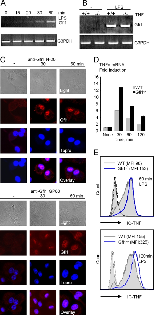
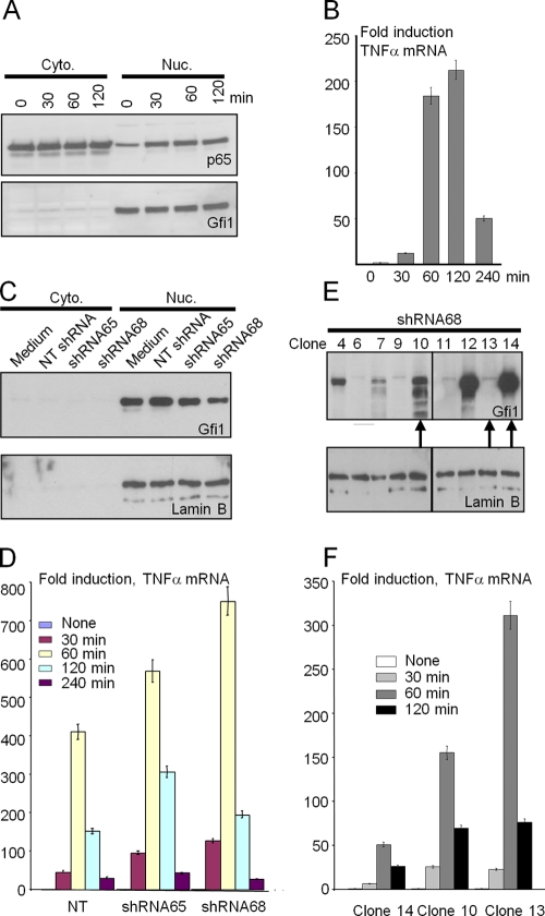
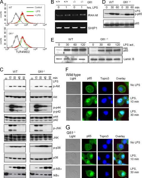
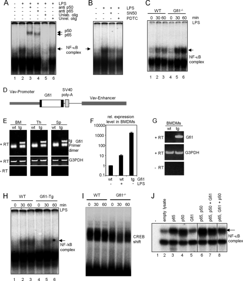
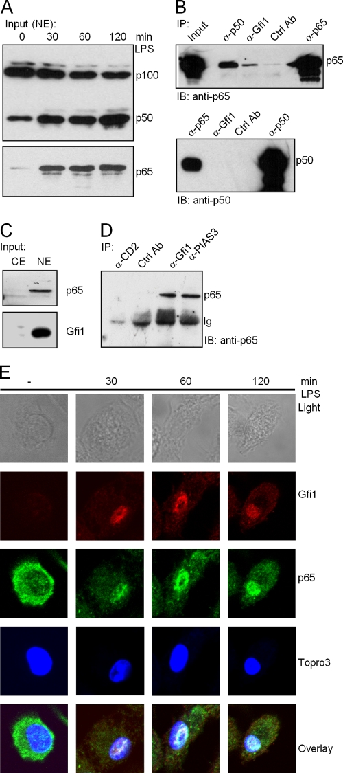
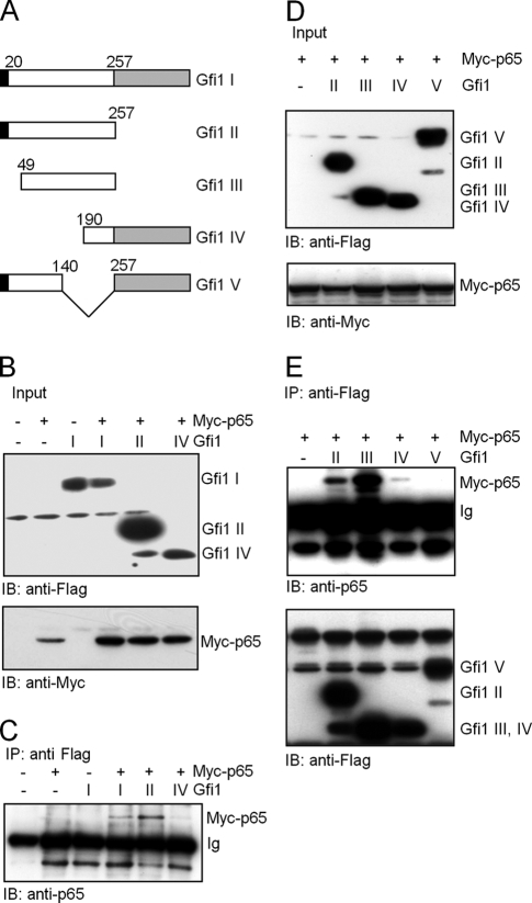
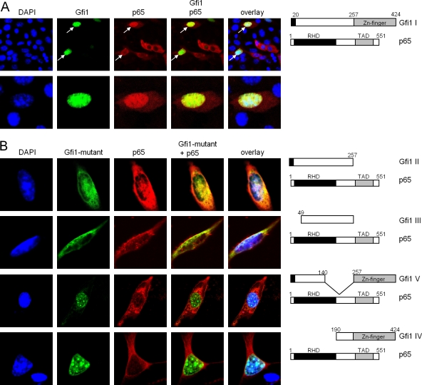
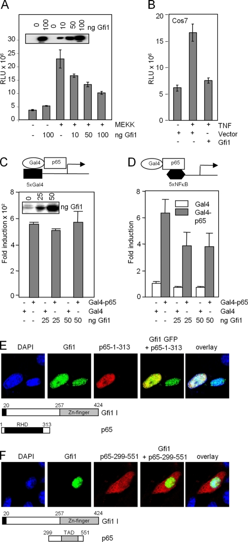
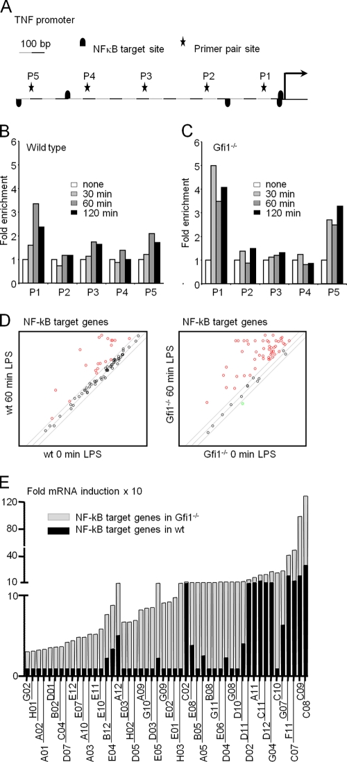
Endotoxin (bacterial lipopolysaccharide [LPS]) causes fatal septic shock via the Toll-like receptor 4 (TLR-4) protein present on innate immunity effector cells, which activates nuclear factor kappa B (NF-kappaB), inducing proinflammatory cytokines, including tumor necrosis factor alpha (TNF-alpha). An early step in this process involves nuclear sequestration of the p65-RelA NF-kappaB subunit, enabling transcriptional activation of target inflammatory cytokine genes. Here, we analyzed the role of the nuclear zinc finger protein Gfi1 in the TLR response using primary bone marrow-derived macrophages. We show that upon LPS stimulation, expression of Gfi1 is induced with kinetics similar to those of nuclear translocation of p65 and that Gfi1 interacts with p65 and inhibits p65-mediated transcriptional transactivation by interfering with p65 binding to target gene promoter DNA. Gfi1-deficient macrophages show abnormally high mRNA levels of the TNF-alpha gene and many other p65 target genes and a higher rate of TNF promoter occupancy by p65 than wild-type cells after LPS stimulation, suggesting that Gfi1 functions as an antagonist of NF-kappaB activity at the level of promoter binding. Our findings identify a new function of Gfi1 as a general negative regulator of the endotoxin-initiated innate immune responses, including septic shock and possibly other severe inflammatory diseases.


Figures

FIG. 1.
FIG. 1.
Induction of Gfi1 expression by TLR4 ligand LPS in wild-type (WT) and Gfi1-deficient (Gfi1/) macrophages. (A) Gfi1 mRNA expression is induced in BMDMs as early as 15 to 30 min after the onset of LPS stimulation (10 ng/ml). (B) RT-PCR analysis of LPS-induced Gfi1 expression in wild-type (+/+) and TNF-deficient (−/−) BMDMs. (C) Wild-type BMDMs were treated with 10 ng/ml of LPS for the indicated times, stained with anti-Gfi1 (left panel, N-20; right panel, GP33), and analyzed using confocal microscopy. (D) TNF-α mRNA is increased in Gfi1-deficient macrophages. Quantification by Q-PCR of Gfi1 mRNA levels using RNA from wild-type (WT) and Gfi1/ BMDMs stimulated with LPS (10 ng/ml) for the indicated times. Representative results from three independent experiments with three independent sets of mice are shown. (E) Flow cytometric analysis of intracellular TNF-α in WT and Gfi1/ BMDMs after LPS treatment for the indicated times. Numbers in brackets represent mean fluorescence intensities. IC-TNF, intracellular TNF-α.
FIG. 2.
FIG. 2.
Regulation of TNF-α transcription by Gfi1 in THP-1 cells. (A) THP-1 cells stimulated with or without LPS (10 ng/ml) for the indicated times. Cytoplasmic (Cyto.) and nuclear (Nuc.) fractions were then blotted against NF-κB p65 and Gfi1 proteins. (B) THP-1 cells were treated as described for panel A, and the induction of TNF-α mRNA was determined by Q-PCR analysis. (C) THP-1 cells were infected with a lentivirus expressing shRNA against Gfi1. Nontargeting (NT) lentivirus was used as a control. Cytoplasmic (Cyto.) and nuclear (Nuc.) fractions were blotted against Gfi1 proteins. (D) Infected cells were stimulated with or without LPS (100 ng/ml) for the indicated times, and the induction of TNF-α mRNA was determined by Q-PCR analysis. (E) Cells infected with lentivirus expressing shRNA68 against Gfi1 were used to single clone THP-1 cells by a limiting dilution assay. Individual clones, indicated by clone number, were used to analyze Gfi1 expression in the nuclear fraction. The arrows at the bottom of the panel indicate the 3 clones chosen for the Q-PCR analysis shown in panel F. (F) Three individual clones (clones 14, 10, and 13) were stimulated with or without LPS (100 ng/ml) for the indicated periods of time, and the induction of TNF-α mRNA was determined by Q-PCR analysis.
FIG. 3.
FIG. 3.
TLR4 signaling is not affected in Gfi1/ BMDMs. Wild-type (WT) and Gfi1/ BMDMs were treated with medium or 10 ng/ml of LPS, and cells were harvested at the indicated time points. (A) Flow cytometry of TLR4-MD2 expression of medium-treated (−LPS) and LPS-stimulated (+LPS) wild-type (WT) and Gfi1/ BMDMs. Staining with irrelevant antibody was used as a control. (B) Expression levels of IRAK-M and SHIP1 determined using RT-PCR. (C) The activation levels of p-Akt, p-Erk, p-JNK, p-p38, and p-IκBα were assessed using immunoblotting. Immunoblot analysis of total endogenous proteins of each signaling molecule was used to ensure equal sample loading. (D) Extracts from WT and Gfi1/ BMDMs treated as indicated were probed with antibodies against NF-κB subunit p65 or the phosphorylated form of p65 (p-p65). (E) Nuclear extracts of wild-type and Gfi1/ BMDMs treated as indicated were probed with antibodies against the NF-κB subunit p65. (F) WT and (G) Gfi1/ BMDMs were treated with 10 ng/ml LPS for the indicated times, and the localization of endogenous NF-κB p65 was assessed by confocal microscopy. Nuclei were visualized by Topro3 staining.
FIG. 4.
FIG. 4.
Enhanced NF-κB DNA binding in Gfi1/ macrophages. Wild-type (WT) and Gfi1/ BMDMs were treated with medium or 10 ng/ml of LPS. Nuclear extracts were collected at the indicated time points for an electrophoretic mobility shift assay (EMSA) using a radiolabeled DNA probe containing an NF-κB (p65-p50) binding site sequence. (A) EMSA with nuclear extracts from WT BMDMs before and after LPS stimulation. An arrow indicates the position of the p65-p50-DNA complex, generated after 1 h of LPS stimulation (arrowheads represent supershift with antibodies recognizing the p65 and p50 subunits of NF-κB). Addition of excess unlabeled NF-κB oligonucleotide or unlabeled irrelevant oligonucleotide (lanes 5 and 6) or NF-κB inhibitors (SN-50 and PDTC) (B) disrupted the formation of the p65-p50-DNA complexes and demonstrated the specificity of the binding reaction. (C) EMSA with nuclear extracts from WT and Gfi1/ BMDMs before and after LPS stimulation. An arrow indicates the induction of the p65-p50-DNA complex after LPS stimulation in wild-type (lanes 1 to 3) and Gfi1/ (lanes 4 to 6) BMDMs. (D) Schematic representation of the vav-Gfi1 construct used to generate transgenic (tg) mice. (E) Expression of the vav-Gfi1 transgene detected by RT-PCR in bone marrow (BM), thymus (Th), and spleen (Sp). (F and G) Expression of the vav-Gfi1 transgene detected in BMDMs by Q-PCR (F) and by RT-PCR (G). (H) EMSA with nuclear BMDM extracts from WT or vav-Gfi1 tg mice before and after LPS stimulation. An arrow indicates the position of the p65-p50-DNA complex, generated after 1 h of LPS stimulation. (I) Control shift experiments with a labeled CREB binding site revealed similar amounts of protein in all samples of nuclear extracts from wild-type (WT) and Gfi1/ BMDMs. (J) EMSA with in vitro-translated proteins as indicated. Lane 7, p65 and p50 were cotranslated, and then Gfi1 in vitro-translated protein was added; lane 8, p65 and Gfi1 were cotranslated, and then p50 in vitro-translated protein was added.
FIG. 5.
FIG. 5.
Endogenous NF-κB subunit p65, but not p50, interacts with Gfi1. (A) Nuclear translocation of NF-κB subunits p65 and p50 after LPS stimulation of THP-1 cells for the indicated times. NE, nuclear extracts. (B) Extracts from THP-1 cells were immunoprecipitated with the indicated antibodies, and the precipitates were analyzed by immunoblotting using anti-p65 antibody (upper panel) or anti-p50 antibody (lower panel). (C) Input levels of endogenous p65 and Gfi1 proteins in Jurkat cells; both are readily detected in Jurkat nuclear extracts (NE) but are absent in cytoplasmic extracts (CE). (D) Extracts from Jurkat cells were immunoprecipitated with the indicated antibodies, and the precipitates were analyzed by immunoblotting using anti-p65 antibodies. Anti-PIAS3 was used as a positive control. Anti-CD2 and anti- atrophrin were used as negative controls and show the specificity of p65 immunoprecipitation. (E) Wild-type BMDMs were treated with 10 ng/ml LPS for the indicated periods of time, and the localization of endogenous NF-κB and Gfi1 proteins was assessed by confocal microscopy. Nuclei were visualized by Topro3 staining.
FIG. 6.
FIG. 6.
Interaction of Gfi1 with the p65 subunit of NF-κB in NIH 3T3 transfected cells. (A) Schematic representation of full-length Gfi1 (I) and four Gfi1 mutants (II, III, IV, and V) used for coimmunoprecipitation after transfection of constructs into NIH 3T3 cells. Gfi1 deletion mutants Gfi1 I (positions 1 to 257) and Gfi1 III (positions 49 to 257) lack the C-terminal zinc finger domain (gray box), and Gfi1 mutants III and IV lack the N-terminal SNAG repressor domain (black box). (B) NIH 3T3 cells were transiently transfected with Flag-tagged full-length Gfi1 (I) or the Gfi1 mutants (II and IV) and Myc-tagged p65, as indicated, and input levels were controlled by immunoblotting (IB) with anti-Myc or anti-Flag antibodies. (C) Whole-cell lysates of the transfected cells were subjected to coimmunoprecipitation (IP) with anti-Flag antibodies, followed by immunoblotting (IB) with anti-p65 antibodies. (D) NIH 3T3 cells were transiently transfected with the indicated Flag-tagged Gfi1 mutants (II, III, IV, and V) and Myc-tagged p65, as indicated, and input levels were controlled by immunoblotting (IB) with anti-Myc or anti-Flag antibodies. (E) Whole-cell lysates of transfected cells were subjected to coimmunoprecipitation (IP) with anti-Flag antibodies, followed by immunoblotting (IB) with anti-p65 or anti-Flag antibodies.
FIG. 7.
FIG. 7.
Gfi1 colocalizes with p65 in the nucleus. (A) NIH 3T3 fibroblasts were transfected with constructs directing the expression of full-length Gfi1 as a fusion protein with GFP or a full-length Myc-tagged p65 protein. Nuclei were visualized by DAPI (4′,6-diamidino-2-phenylindole) staining, and p65 was visualized by staining with anti-Myc antibodies and rhodamine-labeled secondary antibodies. Cells were analyzed with a laser scanning microscope (LSM). The merged pictures demonstrate colocalization of Gfi1 (green) and p65 (red) in cells that coexpress both proteins (white arrows). (B) Cotransfection of the indicated expression constructs, as described for panel A, that allow the production of either full-length Myc-tagged p65 or the Flag-tagged Gfi1 II, III, V, and IV mutants. Gfi1 mutants II and III show a partial colocalization with p65 (first and second rows), but Gfi1 mutants IV and V showed no colocalization (third and fourth rows).
FIG. 8.
FIG. 8.
Gfi1 represses NF-κB transcriptional activity and binds to the p65 Rel homology domain. (A and B) Reporter gene assays with RK13 cells (A) or COS7 cells (B) with a luciferase vector containing 5xNF-κB binding sites. Activation of NF-κB was achieved by cotransfecting the expression constructs for MAP kinase MEKK (A) or by treating the cells with TNF that activates p65 (B). Expression levels of Gfi1 are shown in the inset in panel A. RLU, relative light units. (C and D) Gfi1 interferes with DNA binding and not transcriptional transactivation of p65. Reporter gene assays were performed with NIH 3T3 cells transfected with combinations of expression constructs encoding Gal4 (white bar) or Gal4-p65 (gray bar) fusion proteins, increasing amounts of Flag-Gfi1, and either a 5xGal4 reporter (C) or a 5xNF-κB reporter (D). All data are representative of three independent experiments. (E) Coexpression of the indicated p65 mutants lack-ing either the TAD domain or the Rel homology domain (RHD) with full-length Gfi1. Interaction between Gfi1 and the RHD part of p65 is clearly observed. (F) The p65 mutant containing only the TAD remains in the cytoplasm, indicating that it lacks the ability to interact with Gfi1.
FIG. 9.
FIG. 9.
Enhanced TNF-α promoter occupancy in Gfi1-deficient macrophages and regulation of NF-κB target gene expression by Gfi1. (A) Schematic representation of 1 kb of the TNF-α promoter as assessed by ChIP analysis. Primer pair sites (P1 to P5) and NF-κB target sites are indicated. Wild-type (B) and Gfi1/ (C) BMDMs were treated with medium or 10 ng/ml of LPS for the indicated periods of time. Cells were harvested for ChIP experiments with anti-p65 antibodies, and occupancy of p65 at the indicated sites was determined using Q-PCR with five sets of primer pairs. Data represent the average relative fold inductions at each primer site with respect to the level for nontreated BMDMs (set to 1). The data are representative of two independent sets of experiments. (D) BMDMs from wild-type (wt) and Gfi1/ mice were stimulated with LPS (10 ng/ml) for 60 min or wereleft unstimulated (0 min). Total RNA was extracted and used for the PCR array. Scatter plot of NF-κB target genes induced 60 min after LPS stimulation in BMDMs from WT mice (left panel; red circles represent 19 genes) and Gfi1/ mice (right panel; red circles represent 50 genes). (E) Relative fold mRNA inductions of NF-κB target genes in wild-type mice (WT; black columns) and Gfi1/ mice (KO; gray columns). The code of each NF-κB target gene is indicated based on the manufacturer's indications for the PCR array. The gene products corresponding to the codes are indicated in Table 1.

Similar articles

Cited by

References

    1. Akira, S., and K. Takeda. 2004. Toll-like receptor signalling. Nat. Rev. Immunol. 4:499-511. - PubMed
    1. Akira, S., K. Takeda, and T. Kaisho. 2001. Toll-like receptors: critical proteins linking innate and acquired immunity. Nat. Immunol. 2:675-680. - PubMed
    1. Beutler, B., and E. T. Rietschel. 2003. Innate immune sensing and its roots: the story of endotoxin. Nat. Rev. Immunol. 3:169-176. - PubMed
    1. Blouin, C. C., E. L. Page, G. M. Soucy, and D. E. Richard. 2004. Hypoxic gene activation by lipopolysaccharide in macrophages: implication of hypoxia-inducible factor 1alpha. Blood 103:1124-1130. - PubMed
    1. Burns, K., J. Clatworthy, L. Martin, F. Martinon, C. Plumpton, B. Maschera, A. Lewis, K. Ray, J. Tschopp, and F. Volpe. 2000. Tollip, a new component of the IL-1RI pathway, links IRAK to the IL-1 receptor. Nat. Cell Biol. 2:346-351. - PubMed

Publication types

MeSH terms