. 2010 Aug;30(16):4035-44.
doi: 10.1128/MCB.00175-10. Epub 2010 Jun 14.

Inactivation of Foxo3a and subsequent downregulation of PGC-1 alpha mediate nitric oxide-induced endothelial cell migration

Affiliations

Inactivation of Foxo3a and subsequent downregulation of PGC-1 alpha mediate nitric oxide-induced endothelial cell migration

Sara Borniquel et al. Mol Cell Biol. 2010 Aug.

Abstract

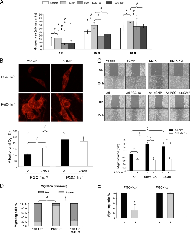
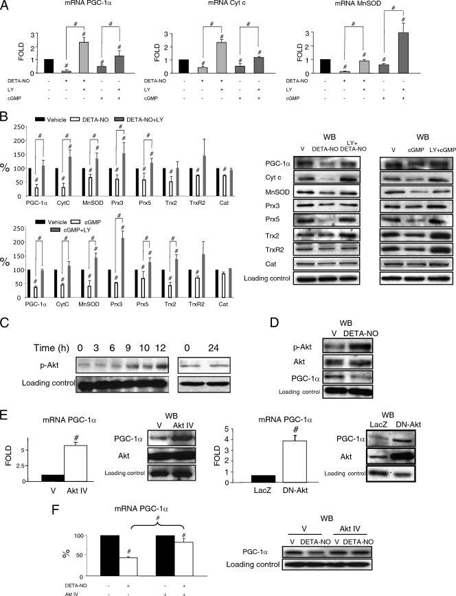
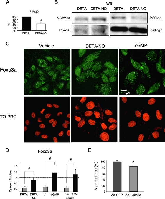
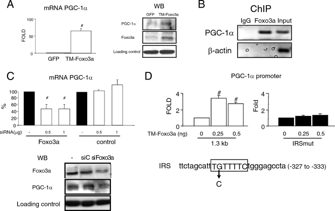
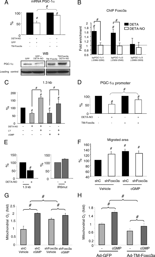
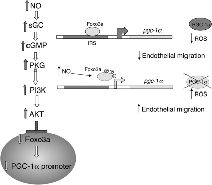
In damaged or proliferating endothelium, production of nitric oxide (NO) from endothelial nitric oxide synthase (eNOS) is associated with elevated levels of reactive oxygen species (ROS), which are necessary for endothelial migration. We aimed to elucidate the mechanism that mediates NO induction of endothelial migration. NO downregulates expression of peroxisome proliferator-activated receptor gamma coactivator 1 alpha (PGC-1 alpha), which positively modulates several genes involved in ROS detoxification. We tested whether NO-induced cell migration requires PGC-1 alpha downregulation and investigated the regulatory pathway involved. PGC-1 alpha negatively regulated NO-dependent endothelial cell migration in vitro, and inactivation of the phosphatidylinositol 3-kinase (PI3K)/protein kinase B (Akt) pathway, which is activated by NO, reduced NO-mediated downregulation of PGC-1 alpha. Expression of constitutively active Foxo3a, a target for Akt-mediated inactivation, reduced NO-dependent PGC-1 alpha downregulation. Foxo3a is also a direct transcriptional regulator of PGC-1 alpha, and we found that a functional FoxO binding site in the PGC-1 alpha promoter is also a NO response element. These results show that NO-mediated downregulation of PGC-1 alpha is necessary for NO-induced endothelial migration and that NO/protein kinase G (PKG)-dependent downregulation of PGC-1 alpha and the ROS detoxification system in endothelial cells are mediated by the PI3K/Akt signaling pathway and subsequent inactivation of the FoxO transcription factor Foxo3a.


Figures

FIG. 1.
FIG. 1.
PGC-1α inhibits endothelial migration. (A) Quantitative analysis of scratch assays in serum-starved confluent BAEC. Cells were preincubated with EUK-189 (100 μM) for 2 h as indicated and then (right after being scratched) incubated with DETA-NO (62 μM) and 8-Br-cGMP (100 μM) as indicated. Repopulation of the denuded area was monitored by confocal microscopy at the indicated times. (B) MitoSOX Red labeling of fixed MLEC, showing mitochondrial superoxide levels in PGC-1α+/+/PGC-1α−/− cells treated with 100 μM 8-Br-cGMP for 6 h. V, vehicle. (C) Scratch wound healing assays with cells infected with PGC-1α or control adenovirus (MOI of 25) at 24 h before the beginning of the assay. Treatments with DETA-NO and 8-Br-cGMP, as shown in panel A, are shown for comparison. Cell migration was monitored at 18 h. (D and E) A total of 10,000 PGC-1α+/+ or PGC-1α−/− MLEC were seeded in the upper chamber of a Transwell insert and allowed to migrate for 18 h before fixing and labeling for cell nuclei. (D) Quantitative analysis of cross sections (z-axis) of Transwell inserts seeded with PGC-1α+/+ or PGC-1α−/− cells, illustrating the distance migrated. Cells were preincubated with EUK-189 (100 μM) for 2 h as indicated. (E) The chart shows the effect of LY294002 (LY; 10 μM) on the Transwell migration of PGC-1α+/+ and PGC-1α−/− MLEC. #, P < 0.05 versus paired control.
FIG. 2.
FIG. 2.
(A and B) NO downregulates PGC-1α and the mitochondrial ROS detoxification system via PKG and PI3K. BAEC were stimulated for 12 h with DETA-NO (62 μM) or 8-Br-cGMP (100 μM) with or without pretreatment with 10 μM LY294002 (LY), as indicated. V, vehicle. (A) mRNA expression levels (quantitative PCR [qPCR]) of PGC-1α and its target genes cytochrome c (CytC) and Mn superoxide dismutase (MnSOD). (B) Western blotting (WB) and band intensity quantification of protein levels of PGC-1α, CytC, MnSOD, peroxiredoxins 3 and 5 (Prx3 and Prx5), thioredoxin 2 (Trx2), thioredoxin reductase 2 (TrxR2), and catalase (Cat). (C to F) Akt is required for NO-induced downregulation of PGC-1α. (C) BAEC were incubated with DETA-NO (62 μM) for 3 to 24 h. Protein levels of phosphorylated Akt (p-Akt) were determined by WB. Zero hours corresponds to untreated cells. (D) WB of Akt, phosphorylated Akt (p-Akt), and PGC-1α in BAEC treated with 62 μM DETA-NO or DETA (V) for 12 h. (E) PGC-1α mRNA expression (quantitative reverse transcription-PCR [qRT-PCR]) and protein levels of PGC-1α (WB) are shown. (Left panels) BAEC were treated with Akt IV (0.5 μM) or vehicle (V) for 12 h. (Right panels) BAEC were infected with dominant negative Akt (DN-Akt) adenovirus or a control (LacZ) at an MOI of 25. (F) BAEC were treated with vehicle (V), DETA-NO (62 μM), Akt IV (0.5 μM), and Akt IV plus DETA-NO for 12 h. PGC-1α mRNA expression (qRT-PCR) and protein levels of PGC-1α (WB) are shown. Control samples were assigned the value of 1. #, P < 0.05 versus paired control.
FIG. 3.
FIG. 3.
NO inactivates Foxo3a. (A) BAEC were transfected with 200 ng (PrFx3X) and, 24 h later, treated with DETA (V) or DETA-NO (62 μM) for 12 h. Control samples were assigned the value 100%. (B) BAEC were incubated with DETA (V) or DETA-NO (62 μM) for 12 h. WB of Foxo3a, phosphorylated Foxo3a (p-Foxo3a) and PGC-1α. c, control. (C and D) Immunofluorescence analysis of the cellular localization of Foxo3a. Serum-starved BAEC were incubated with DETA, DETA-NO (62 μM), or cGMP (100 μM) for 12 h. Cells in 10% FBS were used as a positive control. Nuclei were labeled with TO-PRO-3. (D, left) Distribution of TO-PRO-3 and Foxo3a signal intensity along cross sections of representative cells. (Right) Foxo3a cytosol/nuclear ratio. Averaged staining intensities were calculated from cross-sectional traces. (E) Scratch wound healing assays with cells infected with Foxo3a or control adenovirus (MOI, 10) 24 h before the beginning of the assay. Cell migration was monitored every 2 h for 20 h. #, P < 0.05 versus paired control.
FIG. 4.
FIG. 4.
Foxo3a regulates PGC-1α expression. (A) BAEC were infected with TM-Foxo3a adenovirus (MOI, 10). (Left) PGC-1α mRNA (qPCR); (right) WB of PGC-1α and Foxo3a. GFP, green fluorescent protein. (B) Foxo3a binding to the PGC-1α promoter was determined by ChIP in BAEC. The 18S RNA gene was used as negative control (qPCR; not shown). (C) BAEC were transfected with the indicated amounts of Foxo3a siRNA (siFoxo3a) or control siRNA (siC; GAPDH [glyceraldehyde-3-phosphate dehydrogenase]). (Top) PGC-1α mRNA; (bottom) WB of PGC-1α and Foxo3a. (D) BAEC were cotransfected with 500 ng of the PGC-1α promoter (1.3 kb) or the IRS mutant vector (IRSmut) and the indicated amounts of the TM-Foxo3a vector or the corresponding control. Control samples were assigned the value of 100%. #, P < 0.05 versus paired control.
FIG. 5.
FIG. 5.
NO-induced downregulation of PGC-1α is mediated by Foxo3a. (A) BAEC were infected with TM-Foxo3a (MOI, 10) or control adenovirus and, 24 h later, were incubated with DETA or DETA-NO (62 μM, 12 h) as indicated. (Top) PGC-1α mRNA expression. Control samples (DETA) were assigned the value of 100%. (Bottom) PGC-1α WB. (B) BAEC were incubated with DETA or DETA-NO (62 μM, 12 h), and Foxo3a binding to three positions within the PGC-1α promoter was analyzed by ChIP. IP DNA was analyzed by qPCR. The 18S RNA gene was used as a negative control. (C) BAEC were transfected with 500 ng of the PGC-1α promoter vector and, 24 h later, incubated as indicated with DETA-NO (62 μM), 8-Br-cGMP (100 μM), and LY294002 (10 μM) for 12 h. (D) BAEC were cotransfected with the PGC-1α promoter vector (500 ng) and TM-Foxo3a (1 ng) and treated as indicated with DETA-NO. (E) BAEC were transfected with the PGC-1α promoter or with the IRS mutant vector (500 ng). Control samples (DETA) were assigned the value of 100%. (F) Scratch assay using serum-starved confluent BAEC. Cells were infected with Foxo3a shRNA (shFoxo3a) or control adenovirus (MOI, 25) at 24 h before the beginning of the assay and then treated with 100 μM 8-Br-cGMP as indicated. Cells were allowed to migrate for 18 h. Control samples (control shRNA [shC] plus vehicle) were assigned the value of 100%. (G, H) MitoSOX Red labeling of fixed BAEC infected with shFoxo3a (G), TM-Foxo3a (H), or the corresponding control adenovirus and treated with 100 μM 8-Br-cGMP for 6 h. #, P < 0.05 versus paired control.
FIG. 6.
FIG. 6.
NO-dependent downregulation of PGC-1α expression requires inactivation of Foxo3a by Akt. Foxo3a is a direct and positive regulator of PGC-1α expression, and its inactivation leads to the transcriptional downregulation of PGC-1α. Foxo3a inactivation and PGC-1α downregulation result in reduced ROS detoxification capacity, increased levels of ROS, and induction of endothelial migration.

References

    1. Borniquel, S., I. Valle, S. Cadenas, S. Lamas, and M. Monsalve. 2006. Nitric oxide regulates mitochondrial oxidative stress protection via the transcriptional coactivator PGC-1alpha. FASEB J. 20:1889-1891. - PubMed
    1. Bucci, M., F. Roviezzo, I. Posadas, J. Yu, L. Parente, W. C. Sessa, L. J. Ignarro, and G. Cirino. 2005. Endothelial nitric oxide synthase activation is critical for vascular leakage during acute inflammation in vivo. Proc. Natl. Acad. Sci. U. S. A. 102:904-908. - PMC - PubMed
    1. Daitoku, H., K. Yamagata, H. Matsuzaki, M. Hatta, and A. Fukamizu. 2003. Regulation of PGC-1 promoter activity by protein kinase B and the forkhead transcription factor FKHR. Diabetes 52:642-649. - PubMed
    1. Dimmeler, S., and A. M. Zeiher. 1999. Nitric oxide—an endothelial cell survival factor. Cell Death Differ. 6:964-968. - PubMed
    1. Dudzinski, D. M., J. Igarashi, D. Greif, and T. Michel. 2006. The regulation and pharmacology of endothelial nitric oxide synthase. Annu. Rev. Pharmacol. Toxicol. 46:235-276. - PubMed

Publication types

MeSH terms