. 2010 Aug 20;285(34):26233-44.
doi: 10.1074/jbc.M110.122069. Epub 2010 Jun 14.

Synergism between Wnt3a and heparin enhances osteogenesis via a phosphoinositide 3-kinase/Akt/RUNX2 pathway

Affiliations

Synergism between Wnt3a and heparin enhances osteogenesis via a phosphoinositide 3-kinase/Akt/RUNX2 pathway

Ling Ling et al. J Biol Chem. .

Abstract

A new strategy has emerged to improve healing of bone defects using exogenous glycosaminoglycans by increasing the effectiveness of bone-anabolic growth factors. Wnt ligands play an important role in bone formation. However, their functional interactions with heparan sulfate/heparin have only been investigated in non-osseous tissues. Our study now shows that the osteogenic activity of Wnt3a is cooperatively stimulated through physical interactions with exogenous heparin. N-Sulfation and to a lesser extent O-sulfation of heparin contribute to the physical binding and optimal co-stimulation of Wnt3a. Wnt3a-heparin signaling synergistically increases osteoblast differentiation with minimal effects on cell proliferation. Thus, heparin selectively reduces the effective dose of Wnt3a needed to elicit osteogenic, but not mitogenic responses. Mechanistically, Wnt3a-heparin signaling strongly activates the phosphoinositide 3-kinase/Akt pathway and requires the bone-related transcription factor RUNX2 to stimulate alkaline phosphatase activity, which parallels canonical beta-catenin signaling. Collectively, our findings establish the osteo-inductive potential of a heparin-mediated Wnt3a-phosphoinositide 3-kinase/Akt-RUNX2 signaling network and suggest that heparan sulfate supplementation may selectively reduce the therapeutic doses of peptide factors required to promote bone formation.


Figures

FIGURE 1.
FIGURE 1.
Wnt3a is not sufficient to stimulate ALP activity in MC3T3-E1 cells. A, Wnt3a stimulated LEF1/TCF transcription activity in MC3T3-E1 cells. B, Wnt3a induced nuclear translocation of β-catenin in MC3T3-E1 cells that was assessed by immunofluorescence. Merged images of β-catenin and nuclear 4′,6-diamidino-2-phenylindole (DAPI) staining were shown (green, β-catenin; blue, 4′,6-diamidino-2-phenylindole). Bar, 10 μm. C, Wnt3a stimulated ALP activity in C2C12 but not in MC3T3-E1 cells. Both cells were treated by 50 ng/ml Wnt3a for 72 h before the ALP activity was assayed.
FIGURE 2.
FIGURE 2.
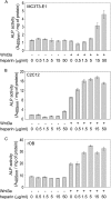
Wnt3a and heparin synergistically enhanced ALP activity. MC3T3-E1 (A) or C2C12 (B) cells seeded at 30,000 cells/cm2 or rOB cells (C) seeded at 5000 cells/cm2 were treated with either 50 ng/ml Wnt3a, increasing doses of heparin (0.5 to 50 μg/ml) or the combination of 50 ng/ml Wnt3a with indicated doses of heparin. Untreated samples served as the control. ALP activity was determined 72 h after treatment.
FIGURE 3.
FIGURE 3.
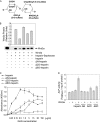
Sulfation on heparin chain is important for its binding affinity and biological activity. A, shown is the structure of disaccharide units of heparin. B, Wnt3a was incubated with heparin-Sepharose in the presence or absence of heparin or desulfated heparin, and the Wnt3a bound to the Sepharose beads was detected by Western blot analysis. The intensity of Wnt3a was quantified by densitometry, and the bar graph plotted was shown above the blot. C, increasing doses of unmodified heparin or desulfated heparin (0.05–150 μg/ml) were immobilized on heparin/GAG binding plates before 50 ng/ml Wnt3a was applied. The amount of Wnt3a bound by these heparins was determined by enzyme-linked immunosorbent assay as described under “Experimental Procedures.” D, MC3T3-E1 cells were stimulated with 50 ng/ml Wnt3a alone or Wnt3a pre-incubated with 50 μg/ml heparin or desulfated heparins for 72 h followed by the determination of ALP activity.
FIGURE 4.
FIGURE 4.
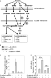
β-Catenin mediated Wnt3a-heparin-stimulated ALP activity. A, shown is an illustration of the interaction of heparin with Wnt and other growth factor signaling pathways that may regulate Wnt dependent osteoblast proliferation and differentiation. IGFBP, IGF-binding protein; BSP, bone sialoprotein; OCN, osteocalcin; OSX, osterix; CKI, cyclin-dependent kinase inhibitor. B, MC3T3-E1 cells were transfected with scramble siRNA or β-catenin siRNA for 24 h. The protein level of β-catenin was assessed by Western blotting. C, cells transfected with β-catenin siRNA or scramble siRNA were further transfected with TOP-FLASH or FOP-FLASH together with Renilla-LUC DNA. 6 h later the cells were exposed to 50 ng/ml Wnt3a for 24 h, and the luciferase activity was measured. D, parallel cultures of β-catenin siRNA-transfected MC3T3-E1 cells were treated with a combination of Wnt3a (50 ng/ml) and heparin (50 μg/ml) for 72 h, and ALP activity assessed.
FIGURE 5.
FIGURE 5.
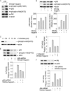
PI3K/Akt pathway is required for Wnt3a-heparin stimulated ALP activity. A and B, p85-PI3K and Akt protein levels as well as their phosphorylation status were measured in MC3T3-E1 cells treated with either 50 ng/ml Wnt3a, 50 μg/ml heparin, or their combination. The relative amount of protein was evaluated by densitometry analysis of the blot in B. C, MC3T3-E1 cells were treated with different concentration of LY294002 for 1 h before the level of phosphorylated Akt (the direct target of activated PI3K) was assessed by Western blotting. D, cells preincubated 1 h with 10 μm LY294002 were stimulated with Wnt3a and heparin for 72 h and then assayed for ALP activity. E and F, MC3T3-E1 cells were transfected with scramble siRNA or siRNA specific for p85 or Akt for 24 h before stimulation with Wnt3a and heparin. The siRNA knock-down efficiency was evaluated by Western blot analysis. ALP activity was measured 72 h post-stimulation.
FIGURE 6.
FIGURE 6.
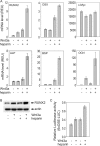
Wnt3a and heparin synergistically increased the expression and activity of osteogenic biomarkers. A, the mRNA levels of several osteogenic transcription factors and osteoblast phenotypic markers were assessed by TaqMan real-time PCR. The samples were prepared in triplicate, and the data represent the mean ± S.E. of three independent experiments. OCN, osteocalcin; OSX, osterix; REU, relative expression unit. B, Western blot analysis of the protein level of RUNX2 after MC3T3-E1 cells were treated with 50 ng/ml Wnt3a, 50 μg/ml heparin, or their combination for 72 h. C, 6xOSE-LUC reporter assay was performed to examine RUNX2 transcription activity as described under “Experimental Procedures.”
FIGURE 7.
FIGURE 7.
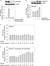
RUNX2 was required for Wnt3a-heparin stimulated ALP activity. A, MC3T3-E1 cells deficient in RUNX2 by RUNX2 siRNA transient transfection were challenged by the combination of Wnt3a and heparin. Then the efficiency of RUNX2 silencing, and the effect on ALP activity was evaluated after 72 h. B, the responsiveness of ALP activity to Wnt3a and heparin was assessed in RUNX2−/− cells. Wild type RUNX2 vector was then re-introduced into RUNX2−/− cells. The efficacy of forced-expression of RUNX2 was evaluated, and the ALP activity was again examined in RUNX2−/− cells treated with Wnt3a and heparin after recovery of RUNX2 expression. C and D, RUNX2−/− cells or RUNX2−/− cells transfected with wild type RUNX2 were seeded at 2500 cells/cm2 and then treated with either 50 ng/ml Wnt3a, increasing doses of heparin (0.5 to 50 μg/ml), or the combination of 50 ng/ml Wnt3a with the indicated doses of heparin. Untreated samples served as controls. ALP activity was determined 72 h after treatment.
FIGURE 8.
FIGURE 8.
PI3K/Akt pathway mediated the effect of Wnt3a and heparin on RUNX2 activity in MC3T3-E1 cells. A, 10 μm LY294002 was applied to the cells 1 h before co-treatment with Wnt3a and heparin. The level of RUNX2 protein was determined 72 h later. B, 24 h after cells were transfected with the specific siRNA against Akt or scramble siRNA, they were stimulated with Wnt3a and heparin for 72 h. The reduced level of Akt and RUNX2 was examined by Western blot analysis. C, cells were transfected with 6xOSE-LUC and Renilla-LUC vectors. After 24 h, cells were challenged with 10 μm LY294002 for 1 h preceding the stimulation with Wnt3a and heparin. The luciferase activity assay was then carried out 48 h later. D, TOP-FLASH or FOP-FLASH together with Renilla-LUC vector was forcibly expressed in cells. 6 h later cells were treated with 10 μm LY294002 for 1 h before the co-treatment of Wnt3a and heparin for another 24 h. The luciferase activity was then determined. The doses of Wnt3a and heparin used above were 50 ng/ml and 50 μg/ml, respectively.
FIGURE 9.
FIGURE 9.
A, shown is a diagram of the signaling mechanism depicting the synergistic activation of ALP by Wnt3a and heparin. B, shown is an illustration of the different role of O-sulfate and N-sulfate groups of heparin in binding to Wnts compared with FGF2. The thickness of the line between the sulfate groups of heparin and the ligand indicates the strength of binding affinity: thinner line, lower affinity; thicker line, higher affinity.

Similar articles

Cited by

References

    1. Guimond S., Maccarana M., Olwin B. B., Lindahl U., Rapraeger A. C. (1993) J. Biol. Chem. 268, 23906–23914 - PubMed
    1. Ishihara M., Shaklee P. N., Yang Z., Liang W., Wei Z., Stack R. J., Holme K. (1994) Glycobiology 4, 451–458 - PubMed
    1. Jackson R. A., Murali S., van Wijnen A. J., Stein G. S., Nurcombe V., Cool S. M. (2007) J. Cell. Physiol. 210, 38–50 - PubMed
    1. Lee J. Y., Choo J. E., Choi Y. S., Lee K. Y., Min D. S., Pi S. H., Seol Y. J., Lee S. J., Jo I. H., Chung C. P., Park Y. J. (2007) J. Biomed Mater. Res. A 83, 970–979 - PubMed
    1. Ling L., Murali S., Dombrowski C., Haupt L. M., Stein G. S., van Wijnen A. J., Nurcombe V., Cool S. M. (2006) J. Cell. Physiol. 209, 811–825 - PubMed

Publication types