

Serum calcium-decreasing factor, caldecrin, inhibits osteoclast differentiation by suppression of NFATc1 activity
- PMID: 20547767
- PMCID: PMC2919108
- DOI: 10.1074/jbc.M109.068742
Serum calcium-decreasing factor, caldecrin, inhibits osteoclast differentiation by suppression of NFATc1 activity
Abstract
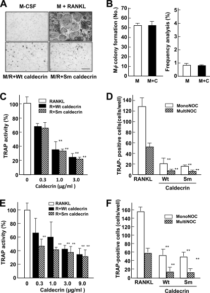
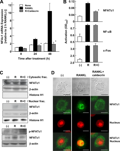
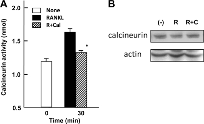
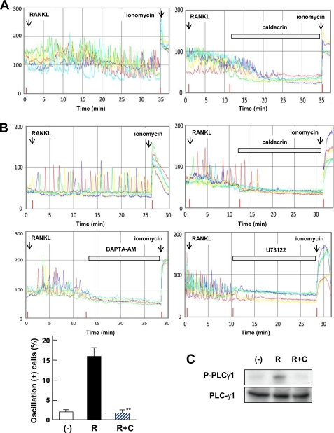
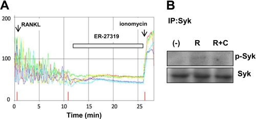
Caldecrin/chymotrypsin C is a novel secretory-type serine protease that was originally isolated as a serum calcium-decreasing factor from the pancreas. Previously, we reported that caldecrin suppressed the bone-resorbing activity of rabbit mature osteoclasts (Tomomura, A., Yamada, H., Fujimoto, K., Inaba, A., and Katoh, S. (2001) FEBS Lett. 508, 454-458). Here, we investigated the effects of caldecrin on mouse osteoclast differentiation induced by macrophage-colony stimulating factor and the receptor activator of NF-kappaB ligand (RANKL) from the monocyte/macrophage cell lineage of bone marrow cells. Wild-type and protease-deficient mutant caldecrin dose-dependently inhibited RANKL-stimulated tartrate-resistant acid phosphatase-positive osteoclast formation from bone marrow cells. Caldecrin did not affect macrophage colony formation from monocyte/macrophage lineage cells or osteoclast progenitor generation in cultures of bone marrow cells. Caldecrin inhibited accumulation of the RANKL-stimulated nuclear factor of activated T-cells, cytoplasmic 1 (NFATc1) mRNA in bone marrow cells, which is a key transcription factor for the differentiation of osteoclasts. Caldecrin also suppressed RANKL-induced differentiation of the RAW264.7 monocyte/macrophage cell line into osteoclasts. Caldecrin reduced the transcriptional activity of NFATc1 in RAW264.7 cells, whereas those of NF-kappaB and c-Fos, which are also transcription factors involved in osteoclast differentiation, were unaffected. Caldecrin inhibited RANKL-stimulated nuclear translocation of NFATc1 and the activity of the calcium/calmodulin-dependent phosphatase, calcineurin. Caldecrin inhibited phospholipase Cgamma1-mediated Ca(2+) oscillation evoked by RANKL stimulation. RANKL-stimulated phosphorylation of spleen tyrosine kinase (Syk) was also attenuated by caldecrin. Taken together, these results indicate that caldecrin inhibits osteoclastogenesis, without its protease activity, by preventing a phospholipase Cgamma1-mediated Ca(2+)oscillation-calcineurin-NFATc1 pathway.
Figures
Similar articles
-
Purification and Biological Function of Caldecrin.Medicines (Basel). 2021 Jul 23;8(8):41. doi: 10.3390/medicines8080041. Medicines (Basel). 2021. PMID: 34436220 Free PMC article. Review.
-
Caldecrin: A pancreas-derived hypocalcemic factor, regulates osteoclast formation and function.World J Biol Chem. 2015 Nov 26;6(4):358-65. doi: 10.4331/wjbc.v6.i4.358. World J Biol Chem. 2015. PMID: 26629319 Free PMC article. Review.
-
Serum calcium-decreasing factor, caldecrin, inhibits receptor activator of NF-κB ligand (RANKL)-mediated Ca2+ signaling and actin ring formation in mature osteoclasts via suppression of Src signaling pathway.J Biol Chem. 2012 May 25;287(22):17963-74. doi: 10.1074/jbc.M112.358796. Epub 2012 Mar 29. J Biol Chem. 2012. PMID: 22461633 Free PMC article.
-
The dectin 1 agonist curdlan regulates osteoclastogenesis by inhibiting nuclear factor of activated T cells cytoplasmic 1 (NFATc1) through Syk kinase.J Biol Chem. 2014 Jul 4;289(27):19191-203. doi: 10.1074/jbc.M114.551416. Epub 2014 May 12. J Biol Chem. 2014. PMID: 24821724 Free PMC article.
-
Zinc inhibits osteoclast differentiation by suppression of Ca2+-Calcineurin-NFATc1 signaling pathway.Cell Commun Signal. 2013 Oct 2;11:74. doi: 10.1186/1478-811X-11-74. Cell Commun Signal. 2013. PMID: 24088289 Free PMC article.
Cited by
-
Nucleosides accelerate inflammatory osteolysis, acting as distinct innate immune activators.J Bone Miner Res. 2011 Aug;26(8):1913-25. doi: 10.1002/jbmr.400. J Bone Miner Res. 2011. PMID: 21472777 Free PMC article.
-
Purification and Biological Function of Caldecrin.Medicines (Basel). 2021 Jul 23;8(8):41. doi: 10.3390/medicines8080041. Medicines (Basel). 2021. PMID: 34436220 Free PMC article. Review.
-
Caldecrin: A pancreas-derived hypocalcemic factor, regulates osteoclast formation and function.World J Biol Chem. 2015 Nov 26;6(4):358-65. doi: 10.4331/wjbc.v6.i4.358. World J Biol Chem. 2015. PMID: 26629319 Free PMC article. Review.
-
Rhinacanthin C Inhibits Osteoclast Differentiation and Bone Resorption: Roles of TRAF6/TAK1/MAPKs/NF-κB/NFATc1 Signaling.PLoS One. 2015 Jun 17;10(6):e0130174. doi: 10.1371/journal.pone.0130174. eCollection 2015. PLoS One. 2015. PMID: 26083531 Free PMC article.
-
Chymotrypsin C (caldecrin) is associated with enamel development.J Dent Res. 2011 Oct;90(10):1228-33. doi: 10.1177/0022034511418231. Epub 2011 Aug 9. J Dent Res. 2011. PMID: 21828354 Free PMC article.
References
-
- Manolagas S. C. (2000) Endocr. Rev. 21, 115–137 - PubMed
-
- Tomomura A., Fukushige T., Noda T., Noikura T., Saheki T. (1992) FEBS Lett. 301, 277–281 - PubMed
-
- Tomomura A., Fukushige T., Tomomura M., Noikura T., Nishii Y., Saheki T. (1993) FEBS Lett. 335, 213–216 - PubMed
-
- Tomomura A., Tomomura M., Fukushige T., Akiyama M., Kubota N., Kumaki K., Nishii Y., Noikura T., Saheki T. (1995) J. Biol. Chem. 270, 30315–30321 - PubMed
-
- Tomomura A., Akiyama M., Itoh H., Yoshino I., Tomomura M., Nishii Y., Noikura T., Saheki T. (1996) FEBS Lett. 386, 26–28 - PubMed
Publication types
MeSH terms
Substances
LinkOut - more resources
Full Text Sources
Miscellaneous
