. 2010 Aug 20;285(34):25987-95.
doi: 10.1074/jbc.M110.100735. Epub 2010 Jun 14.

Vitamin A metabolite, all-trans-retinoic acid, mediates alternative splicing of protein kinase C deltaVIII (PKCdeltaVIII) isoform via splicing factor SC35

Affiliations

Vitamin A metabolite, all-trans-retinoic acid, mediates alternative splicing of protein kinase C deltaVIII (PKCdeltaVIII) isoform via splicing factor SC35

Hercules Apostolatos et al. J Biol Chem. .

Abstract

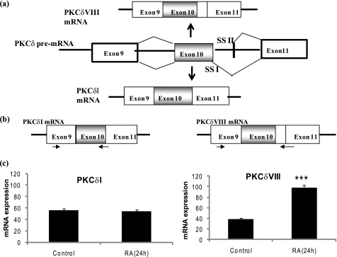
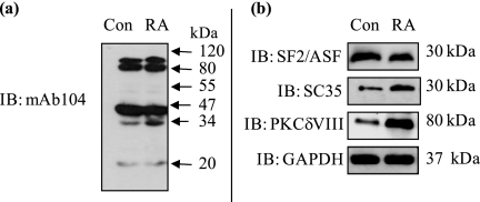
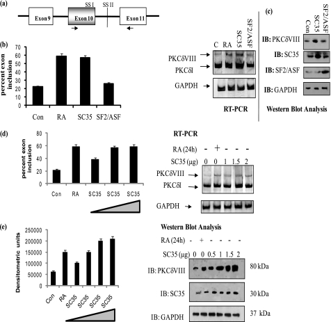
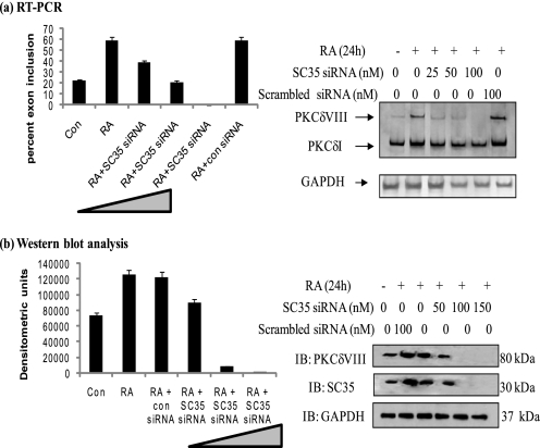
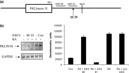
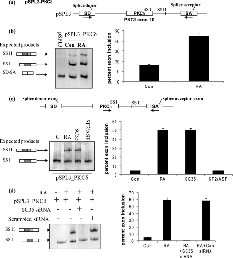
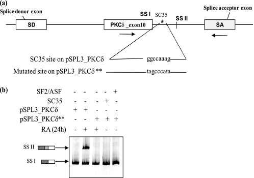
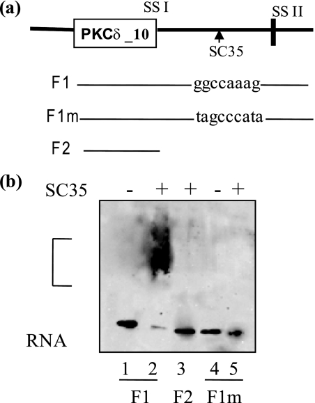
Vitamin A metabolite, all-trans-retinoic acid (RA), induces cell growth, differentiation, and apoptosis and has an emerging role in gene regulation and alternative splicing events. Protein kinase Cdelta (PKCdelta), a serine/threonine kinase, has a role in cell proliferation, differentiation, and apoptosis. We reported an alternatively spliced variant of human PKCdelta, PKCdeltaVIII that functions as a pro-survival protein (1). RA regulates the splicing and expression of PKCdeltaVIII via utilization of a downstream 5' splice site of exon 10 on PKCdelta pre-mRNA. Here, we further elucidate the molecular mechanisms involved in RA regulation of alternative splicing of PKCdeltaVIII mRNA. Overexpression and knockdown of the splicing factor SC35 (i.e. SRp30b) indicated that it is involved in PKCdeltaVIII alternative splicing. To identify the cis-elements involved in 5' splice site selection we cloned a minigene, which included PKCdelta exon 10 and its flanking introns in the pSPL3 splicing vector. Alternative 5' splice site utilization in the minigene was promoted by RA. Further, co-transfection of SC35 with PKCdelta minigene promoted selection of 5' splice site II. Mutation of the SC35 binding site in the PKCdelta minigene abolished RA-mediated utilization of 5' splice splice II. RNA binding assays demonstrated that the enhancer element downstream of PKCdelta exon 10 is a SC35 cis-element. We conclude that SC35 is pivotal in RA-mediated PKCdelta pre-mRNA alternative splicing. This study demonstrates how a nutrient, vitamin A, via its metabolite RA, regulates alternative splicing and thereby gene expression of the pro-survival protein PKCdeltaVIII.


Figures

FIGURE 1.
FIGURE 1.
a, schematic of alternative 5′ splice site selection in human PKCδ pre-mRNA exon 10 that results in the generation of PKCδI mRNA and PKCδVIII mRNA, which differ by 93 bp in the V3 hinge region. RA promotes expression of PKCδVIII mRNA. SSI: 5′ splice site I; SSII: 5′ splice site II. b, schematic of the primers specific for PKCδI and PKCδVIII used in real time RT-PCR such that they span the exon-exon boundaries. c, primary human neuronal cells from hippocampus were treated with or without RA (10 μm) for 24 h. Total RNA was extracted, and real time RT-PCR analysis using SYBR green was performed in triplicate and repeated three times in separate experiments. The absolute mRNA expression of PKCδI and PKCδVIII transcripts normalized to GAPDH are shown. PKCδVIII expression increases significantly following 24 h of RA treatment; ***, p < 0.0001 (by two-tailed Student's t test).
FIGURE 2.
FIGURE 2.
Detection of SR proteins involved in RA-mediated PKCδVIII expression. NT2 cells were treated with RA (10 μm) for 24 h or without RA (control), and Western blot analysis was performed on whole cell lysates using (a) mAb104 antibody that detects all SR proteins and (b) specific antibodies as indicated in the figure. Molecular masses are indicated (kDa). Gels are representative of three separate experiments, and results indicate that SC35 may be involved in increased expression of PKCδVIII by RA. Results demonstrate an increase in SC35 levels concurrent with an increase in PKCδVIII expression upon RA treatment.
FIGURE 3.
FIGURE 3.
SC35 but not SF2/ASF promotes PKCδVIII expression. a, schematic of primer positions used in PCR amplification. These primers detect PKCδI and PKCδVIII simultaneously. b, NT2 cells were transfected with 2 μg of SC35 or SF2/ASF or treated with RA (10 μm) for 24 h. Total RNA was extracted, and RT-PCR was performed using human PKCδ primers as shown above. 5% of the products were separated by PAGE and silver stained for visualization. The graph represents percent exon inclusion calculated as PKCδVIII/(δVIII + δI) × 100 in these samples and is representative of mean ± S.E. in three experiments. c, whole cell lysates were extracted from NT2 cells transfected with 2 μg of SC35 or SF2/ASF. Western blot analysis was performed using specific antibodies as indicated in the figure. The experiments were repeated three times with similar results. d, increasing amounts of SC35 (0 to 2 μg) were transfected into NT2 cells and treated with or without RA (10 μm, 24 h). Total RNA was extracted and RT-PCR was performed using human PKCδ primers as shown above. 5% of the products were separated by PAGE and silver stained for visualization. Graph represents percent exon inclusion calculated as PKCδVIII/(δVIII + δI) × 100 in these samples and is representative of mean ± S.E. in three experiments. e, simultaneously, Western blot analysis was performed on whole cell lysates extracted from NT2 cells transfected with 0–2 μg of SC35, using antibodies as indicated within the figure. The graph represents four experiments performed separately and represents PKCδVIII densitometric units normalized to GAPDH as mean ± S.E. The triangle in the graphs indicates increasing amounts of SC35. Results indicate that SC35 promotes PKCδVIII expression in a dose-dependent manner thereby mimicking the RA response.
FIGURE 4.
FIGURE 4.
Knockdown of SC35 inhibits RA-mediated increased expression of PKCδVIII. Increasing amounts of SC35 siRNA (0–150 nm) were transfected into NT2 cells. Scrambled siRNA was used as a control (con siRNA). Post-transfection, cells were treated with or without RA (10 μm, 24 h). a, total RNA was extracted, and RT-PCR was performed using human PKCδ primers as shown above. 5% of the products were separated by PAGE and silver stained for visualization. Graph represents percent exon inclusion calculated as PKCδVIII/(δVIII + δI) × 100 in these samples and is representative of mean ± S.E. in three experiments. b, simultaneously, whole cell lysates were collected, and Western blot analysis was performed using antibodies as indicated. Graph represents four experiments performed separately and expressed as mean ± S.E. of densitometric units. The triangle in the graphs indicates increasing amounts of SC35 siRNA. Results indicate that knockdown of SC35 inhibits RA-mediated increased expression of PKCδVIII.
FIGURE 5.
FIGURE 5.
Analysis of putative cis-elements and ASO. a, schematic of position of ASOs on PKCδ pre-mRNA. The putative SC35 cis-element lies between 5′ splice site I and II of PKCδ exon 10. SSI: 5′ splice site I; SSII: 5′ splice site II. b, ASOs were transfected into NT2 cells and after overnight incubation cells were treated with or without RA (10 μm, 24 h). The gel represents experiments conducted with scrambled ASO (control), ASO 81 (corresponding to putative SC35 binding site) and ASO 80, which is in close proximity to ASO81. Total RNA was extracted and RT-PCR performed using PKCδVIII-specific primers. 5% products were separated on PAGE and detected by silver nitrate staining. The graph indicates PKCδVIII densitometric units normalized to GAPDH and is representative of mean ± S.E. in three separate experiments. Results indicate that ASO81, which corresponds to the putative SC35 cis-element, inhibits RA-mediated increased expression of PKCδVIII.
FIGURE 6.
FIGURE 6.
Minigene analysis demonstrates that RA promotes utilization of 5′ splice site II on PKCδ exon 10 pre-mRNA. a, schematic represents PKCδ pre-mRNA exon 10 and flanking introns cloned into pSPL3 splicing vector between the SD and SA exons. The resulting minigene is referred to as pSPL3_PKCδ minigene. Arrows indicate position of primers used in RT-PCR analysis. b, pSPL3_PKCδ minigene and pSPL3 empty vector were transfected overnight, and then the cells were treated with or without 10 μm RA for 24 h. Total RNA was extracted and RT-PCR performed using primers as described above. Expected products are SD-SA: constitutive splicing; SSI: usage of 5′ splice site I; SSII: usage of 5′ splice site II. c, 2 μg of SC35 or SF2/ASF was co-transfected along with the pSPL3_PKCδ splicing minigene. In separate wells, 10 μm RA was added for 24 h. Total RNA was extracted and RT-PCR performed using PKCδ exon 10 and SA primers as shown in the schematic. SSI: usage of 5′ splice site I; SSII: usage of 5′ splice site II. d, SC35 siRNA (100 nm) or scrambled siRNA was co-transfected with pSPL3_PKCδ minigene. 10 μm RA was added to wells as indicated. Total RNA was extracted and RT-PCR performed using PKCδ exon 10 and SA primers as shown above in c. 5% of the products were separated by PAGE and silver stained for visualization. Graphs represent percent exon inclusion calculated as SS II/(SS II + SSI) × 100 in the samples and are representative of four experiments performed separately. These results demonstrate that co-transfection of SC35 with the pSPL3_PKCδ minigene promotes utilization of 5′ splice site II. Further, RA is unable to promote utilization of 5′ splice site II on PKCδVIII pre-mRNA in the absence of SC35.
FIGURE 7.
FIGURE 7.
Mutation of putative SC35 binding site inhibits RA-mediated utilization of 5′ splice site II utilization on the minigene. a, schematic of the position and sequence of the putative SC35 cis-element on the pSPL3_PKCδ splicing minigene. Arrows indicate the position of primers used in PCR analysis. Putative SC35 binding site ggccaaag was mutated to tagcccaga on the minigene. b, resulting mutated minigene pSPL3_PKCδ** was transfected into NT2 cells. In separate wells, the mutated minigene pSPL3_PKCδ** was co-transfected with either 2 μg of SC35 or SF2/ASF. The original pSPL3_PKCδ splicing minigene was also transfected in a separate well. After overnight transfection, NT2 cells were treated with or without 10 μm RA for 24 h. Total RNA was extracted and RT-PCR performed using primers for PKCδ exon 10 sense and SA antisense as shown. 5% of the products were separated by PAGE and silver stained for visualization. SSI: usage of 5′ splice site I; SSII: usage of 5′ splice site II. Graph represents percent exon inclusion calculated as SS II/(SS II + SSI) × 100 and is representative of three experiments performed separately. Results indicate that mutation of the enhancer element ggccaaag abolishes the ability of RA or SC35 to promote utilization of 5′ splice site II on PKCδ splicing minigene.
FIGURE 8.
FIGURE 8.
Gel mobility assays of F1 and mutated F1 with purified recombinant SC35. a, schematic representation of the position of PKCδ transcripts F1, F1m and F2 used in the gel binding assays. F1 contains exon 10 and 120 bp of flanking 5′ sequence, which includes the enhancer sequence ggccaaag; schematic also indicates its position on the PKCδ pre-mRNA. F1m is the same as F1 with the enhancer sequence mutated to tagcccata. F2 transcript contains PKCδ10 exon only. b, the biotin-labeled in vitro transcribed RNA sequences were incubated with recombinant SC35 at 30 °C for 20 min. The complex was run on an 8% polyacrylamide gel and transferred to a nylon membrane. Western blot analysis was performed using an avidin-HRP conjugate. Lanes are 1: F1; 2: F1 +SC35; 3: F2 +SC35; 4: F1m; 5: F1m +SC35. The bracket indicates RNA-protein complex. The gel represents four experiments performed separately. Results indicate that ggccaaag is an SC35 cis-element on PKCδ pre-mRNA.

Similar articles

Cited by

References

    1. Jiang K., Apostolatos A. H., Ghansah T., Watson J. E., Vickers T., Cooper D. R., Epling-Burnette P. K., Patel N. A. (2008) Biochemistry 47, 787–797 - PubMed
    1. Nishizuka Y. (1986) Science 233, 305–312 - PubMed
    1. Emoto Y., Manome Y., Meinhardt G., Kisaki H., Kharbanda S., Robertson M., Ghayur T., Wong W. W., Kamen R., Weichselbaum R. (1995) EMBO J. 14, 6148–6156 - PMC - PubMed
    1. Ghayur T., Hugunin M., Talanian R. V., Ratnofsky S., Quinlan C., Emoto Y., Pandey P., Datta R., Huang Y., Kharbanda S., Allen H., Kamen R., Wong W., Kufe D. (1996) J. Exp. Med. 184, 2399–2404 - PMC - PubMed
    1. Kohtz J. D., Jamison S. F., Will C. L., Zuo P., Lührmann R., Barcia-Blanco M. A., Manley J. L. (1994) Nature 368, 119–124 - PubMed

Publication types