

Proteomic identification of phosphatidylinositol (3,4,5) triphosphate-binding proteins in Dictyostelium discoideum
- PMID: 20547830
- PMCID: PMC2900710
- DOI: 10.1073/pnas.1006153107
Proteomic identification of phosphatidylinositol (3,4,5) triphosphate-binding proteins in Dictyostelium discoideum
Abstract
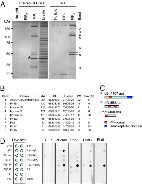
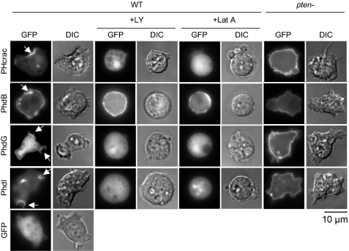
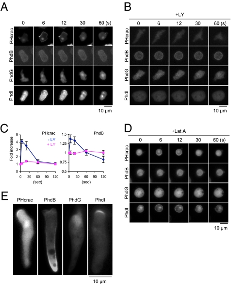
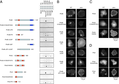
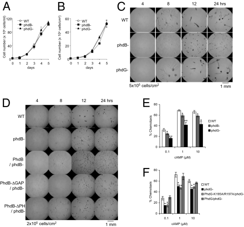
Phosphatidylinositol (3,4,5)-triphosphate (PtdInsP(3)) mediates intracellular signaling for directional sensing and pseudopod extension at the leading edge of migrating cells during chemotaxis. How this PtdInsP(3) signal is translated into remodeling of the actin cytoskeleton is poorly understood. Here, using a proteomics approach, we identified multiple PtdInsP(3)-binding proteins in Dictyostelium discoideum, including five pleckstrin homology (PH) domain-containing proteins. Two of these, the serine/threonine kinase Akt/protein kinase B and the PH domain-containing protein PhdA, were previously characterized as PtdInsP(3)-binding proteins. In addition, PhdB, PhdG, and PhdI were identified as previously undescribed PH domain-containing proteins. Specific PtdInsP(3) interactions with PhdB, PhdG, and PhdI were confirmed using an in vitro lipid-binding assay. In cells, PhdI associated with the plasma membrane in a manner dependent on both the PH domain and PtdInsP(3). Consistent with this finding, PhdI located to the leading edge in migrating cells. In contrast, PhdG was found in the cytosol in WT cells. However, when PtdInsP(3) was overproduced in pten(-) cells, PhdG located to the plasma membrane, suggesting its weak affinity for PtdInsP(3). PhdB was found to bind to the plasma membrane via both PtdInsP(3)-dependent and -independent mechanisms. The PtdInsP(3)-independent interaction was mediated by the middle domain, independent of the PH domain. In migrating cells, the majority of PhdB was found at the lagging edge. Finally, we deleted the genes encoding PhdB and PhdG and demonstrated that both proteins are required for efficient chemotaxis. Thus, this study advances our understanding of the PtdInsP(3)-mediated signaling mechanisms that control directed cell migration in chemotaxis.
Conflict of interest statement
The authors declare no conflict of interest.
Figures
Similar articles
-
Role of phosphatidylinositol 3' kinase and a downstream pleckstrin homology domain-containing protein in controlling chemotaxis in dictyostelium.J Cell Biol. 2001 May 14;153(4):795-810. doi: 10.1083/jcb.153.4.795. J Cell Biol. 2001. PMID: 11352940 Free PMC article.
-
Sensitization of Dictyostelium chemotaxis by phosphoinositide-3-kinase-mediated self-organizing signalling patches.J Cell Sci. 2004 Jun 15;117(Pt 14):2925-35. doi: 10.1242/jcs.01143. Epub 2004 May 25. J Cell Sci. 2004. PMID: 15161938
-
Single-molecule analysis of chemoattractant-stimulated membrane recruitment of a PH-domain-containing protein.J Cell Sci. 2006 Mar 15;119(Pt 6):1071-9. doi: 10.1242/jcs.02824. Epub 2006 Feb 28. J Cell Sci. 2006. PMID: 16507590
-
Leading the way: Directional sensing through phosphatidylinositol 3-kinase and other signaling pathways.J Cell Sci. 2003 Sep 1;116(Pt 17):3471-8. doi: 10.1242/jcs.00703. J Cell Sci. 2003. PMID: 12893811 Review.
-
Moving towards a paradigm: common mechanisms of chemotactic signaling in Dictyostelium and mammalian leukocytes.Cell Mol Life Sci. 2014 Oct;71(19):3711-47. doi: 10.1007/s00018-014-1638-8. Epub 2014 May 21. Cell Mol Life Sci. 2014. PMID: 24846395 Free PMC article. Review.
Cited by
-
Characterization of PTEN mutations in brain cancer reveals that pten mono-ubiquitination promotes protein stability and nuclear localization.Oncogene. 2017 Jun 29;36(26):3673-3685. doi: 10.1038/onc.2016.493. Epub 2017 Mar 6. Oncogene. 2017. PMID: 28263967 Free PMC article.
-
A new class of cancer-associated PTEN mutations defined by membrane translocation defects.Oncogene. 2015 Jul;34(28):3737-43. doi: 10.1038/onc.2014.293. Epub 2014 Sep 29. Oncogene. 2015. PMID: 25263454 Free PMC article.
-
Comprehensive Identification of Lipid-Membrane Protein Interactions via Advanced Proteomics and Extended Lipid-Immobilized Bead Technology.Anal Chem. 2025 Apr 29;97(16):8880-8889. doi: 10.1021/acs.analchem.5c00074. Epub 2025 Apr 15. Anal Chem. 2025. PMID: 40233011 Free PMC article.
-
Mechanism of human PTEN localization revealed by heterologous expression in Dictyostelium.Oncogene. 2014 Dec 11;33(50):5688-96. doi: 10.1038/onc.2013.507. Epub 2013 Dec 2. Oncogene. 2014. PMID: 24292679 Free PMC article.
-
YPIBP: A repository for phosphoinositide-binding proteins in yeast.Comput Struct Biotechnol J. 2021 Jun 24;19:3692-3707. doi: 10.1016/j.csbj.2021.06.035. eCollection 2021. Comput Struct Biotechnol J. 2021. PMID: 34285772 Free PMC article.
References
-
- Iijima M, Huang YE, Devreotes P. Temporal and spatial regulation of chemotaxis. Dev Cell. 2002;3:469–478. - PubMed
-
- Van Haastert PJ, Devreotes PN. Chemotaxis: Signalling the way forward. Nat Rev Mol Cell Biol. 2004;5:626–634. - PubMed
-
- Fey P, Kowal AS, Gaudet P, Pilcher KE, Chisholm RL. Protocols for growth and development of Dictyostelium discoideum. Nat Protoc. 2007;2:1307–1316. - PubMed
-
- Willard SS, Devreotes PN. Signaling pathways mediating chemotaxis in the social amoeba, Dictyostelium discoideum. Eur J Cell Biol. 2006;85:897–904. - PubMed
-
- Chung CY, Firtel RA. Signaling pathways at the leading edge of chemotaxing cells. J Muscle Res Cell Motil. 2002;23:773–779. - PubMed
Publication types
MeSH terms
Substances
Grants and funding
LinkOut - more resources
Full Text Sources
Other Literature Sources
Molecular Biology Databases
Research Materials
