. 2010 Jun 29;107(26):11853-8.
doi: 10.1073/pnas.1003317107. Epub 2010 Jun 14.

Fgf-9 is required for angiogenesis and osteogenesis in long bone repair

Affiliations

Fgf-9 is required for angiogenesis and osteogenesis in long bone repair

Björn Behr et al. Proc Natl Acad Sci U S A. .

Abstract

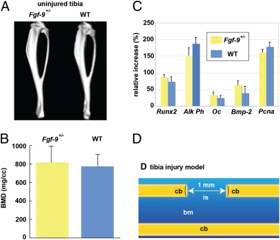
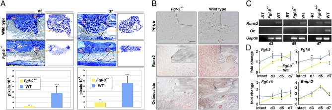
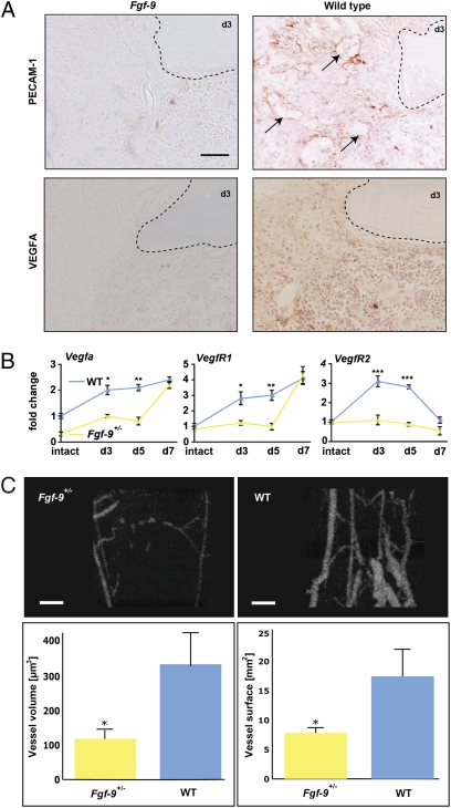
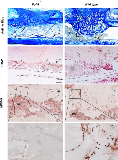
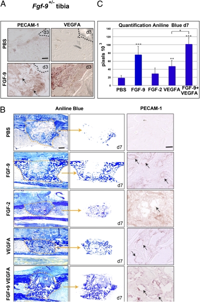
Bone healing requires a complex interaction of growth factors that establishes an environment for efficient bone regeneration. Among these, FGFs have been considered important for intrinsic bone-healing capacity. In this study, we analyzed the role of Fgf-9 in long bone repair. One-millimeter unicortical defects were created in tibias of Fgf-9(+/-) and wild-type mice. Histomorphometry revealed that half-dose gene of Fgf-9 markedly reduced bone regeneration as compared with wild-type. Both immunohistochemistry and RT-PCR analysis revealed markedly decreased levels of proliferating cell nuclear antigen (PCNA), Runt-related transcription factor 2 (Runx2), osteocalcin, Vega-a, and platelet endothelial cell adhesion molecule 1 (PECAM-1) in Fgf-9(+/-) defects. muCT angiography indicated dramatic impairment of neovascularization in Fgf-9(+/-) mice as compared with controls. Treatment with FGF-9 protein promoted angiogenesis and successfully rescued the healing capacity of Fgf-9(+/-) mice. Importantly, although other pro-osteogenic factors [Fgf-2, Fgf-18, and bone morphogenic protein 2 (Bmp-2)] still were present in Fgf-9(+/-) mice, they could not compensate for the haploinsufficiency of the Fgf-9 gene. Therefore, endogenous Fgf-9 seems to play an important role in long bone repair. Taken together our data suggest a unique role for Fgf-9 in bone healing, presumably by initiating angiogenesis through Vegf-a. Moreover, this study further supports the embryonic phenotype previously observed in the developing limb, thus promoting the concept that healing processes in adult organisms may recapitulate embryonic skeletal development.


Conflict of interest statement

The authors declare no conflict of interest.

Figures

Fig. 1.
Fig. 1.
(A) CT scans of tibia in Fgf-9+/− and wild-type mice showed no apparent differences in phenotype. (B) Bone marrow density (BMD) measurements were in the same range, and no difference could be observed. (C) Real-time PCR analysis did not reveal significant differences in the expression profile of osteogenic and proliferative markers. (D) Model of tibia injury. Unicortical tibia injuries were performed by drilling a 1-mm hole in one cortex, leaving the opposite cortex intact. Alk ph, alkaline phosphatase; bm, bone marrow; cb, cortical bone; is, injury site; Oc, osteocalcin.
Fig. 2.
Fig. 2.
Bone healing was impaired in Fgf-9+/− mice. (A) (Upper) Aniline blue staining of unicortical tibial defects at days 5 (Left) and 7 (Right). At both time points, bone regeneration was markedly reduced in Fgf-9+/− mice compared with wild-type mice. The injury site is marked in orange and is segregated in the Insets. (Lower) The amount of new bone formation was quantified with histomorphometry performed on aniline blue-stained slides, revealing a remarkable drop in bone regeneration in Fgf-9+/− mice. ***P < 0.0005. (B) Immunohistochemistry for PCNA, Runx2, and osteocalcin in Fgf-9+/− and wild-type mice. PCNA staining revealed fewer proliferating cells in Fgf-9+/− mice. For Runx2, only faint staining was observed at day 3 in defects of Fgf-9+/− mice, whereas strong nuclear staining was observed in defects in wild-type mice. At day 7, no staining for osteocalcin was observed in Fgf-9+/− mice, whereas staining was observed in wild-type mice. Dashed lines indicate the cortical bone. (Scale bars, 200 μm in A and 50 μm in B.) (C) RT-PCR analysis of Runx2 and osteocalcin (Oc) performed on regenerating bone tissue harvested from Fgf-9+/− and wild-type mice. (D) Real-time PCR analysis for Fgf-2, Fgf-9, Fgf-18, and Bmp-2. *P < 0.05, **P < 0.005 calculated using Student's t test.
Fig. 3.
Fig. 3.
Neovascularization was impaired in bone regeneration of Fgf-9+/− mice. (A) Immunohistochemistry for PECAM-1 and VEGF-A in Fgf-9+/− and wild-type mice. At day 3, only faint PECAM-1 staining was observed in defects of Fgf-9+/− mice, whereas intense PECAM-1 staining was detected in wild-type mice. Arrows indicate blood vessels. Immunohistochemistry for VEGF-A showed less staining in Fgf-9+/− than in wild-type mice at day 3. Dashed lines indicate cortical bone. (B) Real-time PCR time-course analysis of regenerating bone tissue from Fgf-9+/− and wild-type mice for Vegf-a, VegfR1, and VegfR2. *P < 0.05; **P < 0.005; ***P < 0.0005 calculated using Student's t test. (C) μCT angiography of defects in Fgf-9+/− and wild-type mice at day 7. Angiography revealed impaired neovascularization in defects of Fgf-9+/− mice. The histogram shows that both vessel volume and surface were decreased in defects of Fgf-9+/− mice. *P < 0.05. (Scale bars, 50 μm in A and 200 μm in C.)
Fig. 4.
Fig. 4.
Osteoclastogenesis was impaired in Fgf-9+/− mice. Bone regeneration was impaired in Fgf-9+/− compared with wild-type mice at day 7 (Top row, aniline blue staining,). On adjacent sections, faint TRAP staining was observed in defects of Fgf-9+/− compared with wild-type mice at day 7 (Second row). Immunohistochemistry for MMP-9 revealed less staining in the regenerating bone of Fgf-9+/− mice than in wild-type mice (Third row). Boxed areas are enlarged in the bottom row. Dashed lines indicate the cortical bone. Arrowheads indicate MMP-9–positive cells. bm, bone marrow. (Scale bars, 200 μm for aniline blue and TRAP staining and 100 μm for MMP-9 staining.)
Fig. 5.
Fig. 5.
Defects in Fgf-9+/− mice can be rescued with FGF-9 and VEGF-A. (A) PECAM-1 and VEGF-A immunohistochemistry of defects in Fgf-9+/− mice treated with PBS or 2 μg FGF-9 revealed increased vessel formation and VEGF-A staining in the FGF-9–treated defects at day 3. (B) Aniline blue staining of defects in Fgf-9+/− mice treated with PBS, 2 μg FGF-9, 2 μg FGF-2, 2 μg VEGF-A, or a combination of 2 μg FGF-9 and 2 μg VEGF-A. The injury site is marked in orange (Left column) and is segregated in the middle column. Accompanying PECAM-1 staining performed at day 7 revealed vessel formation (arrows) in the treated groups as compared with the PBS control (Right column). (C) Histomorphometry revealed rescue of the defects with all treatments, but FGF-9 and FGF-9 in combination with VEGF-A were the most efficient. *P < 0.05 for combined FGF-9 and VEGF-A treatment vs. VEGF-A treatment alone; **P < 0.005 and ***P < 0.0005 for treatment groups vs. PBS. (Scale bars, for PECAM and 200 μm for aniline blue.)

Similar articles

Cited by

References

    1. Einhorn TA. The cell and molecular biology of fracture healing. Clin Orthop Relat Res. 1998;46(355, Suppl):S7–S21. - PubMed
    1. Gerstenfeld LC, Cullinane DM, Barnes GL, Graves DT, Einhorn TA. Fracture healing as a post-natal developmental process: Molecular, spatial, and temporal aspects of its regulation. J Cell Biochem. 2003;88:873–884. - PubMed
    1. Schindeler A, McDonald MM, Bokko P, Little DG. Bone remodeling during fracture repair: The cellular picture. Semin Cell Dev Biol. 2008;19:459–466. - PubMed
    1. Eswarakumar VP, et al. The IIIc alternative of Fgfr2 is a positive regulator of bone formation. Development. 2002;129:3783–3793. - PubMed
    1. Deng C, Wynshaw-Boris A, Zhou F, Kuo A, Leder P. Fibroblast growth factor receptor 3 is a negative regulator of bone growth. Cell. 1996;84:911–921. - PubMed

Publication types

MeSH terms

Substances

LinkOut - more resources