. 2010 Jun 29;107(26):11835-40.
doi: 10.1073/pnas.0914569107. Epub 2010 Jun 14.

Parkin overexpression selects against a deleterious mtDNA mutation in heteroplasmic cybrid cells

Affiliations

Parkin overexpression selects against a deleterious mtDNA mutation in heteroplasmic cybrid cells

Der-Fen Suen et al. Proc Natl Acad Sci U S A. .

Abstract

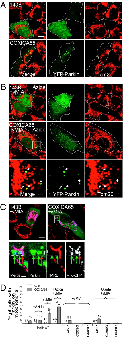
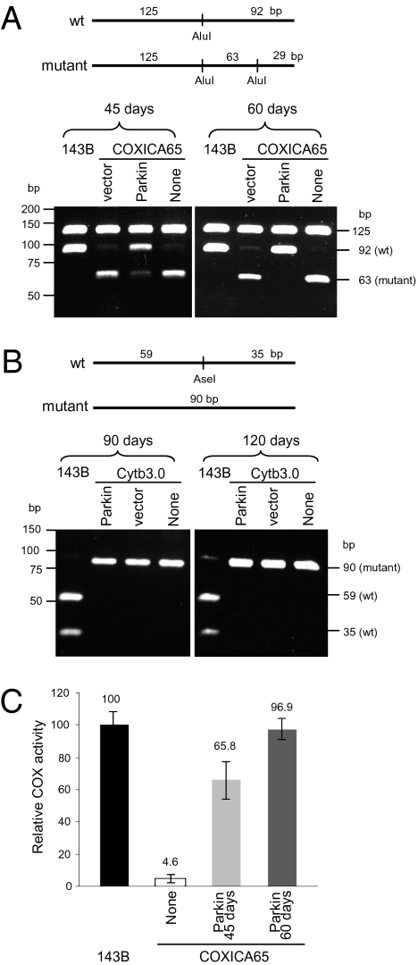
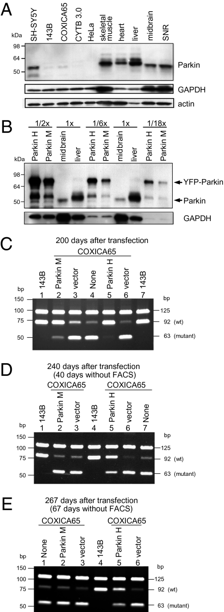
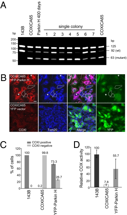
Mitochondrial genomes with deleterious mutations can replicate in cells along with wild-type genomes in a state of heteroplasmy, and are a cause of severe inherited syndromes, such as mitochondrial myopathy, encephalopathy, lactic acidosis, and stroke (MELAS), neuropathy, ataxia, retinitis pigmentosa-maternally inherited Leigh syndrome (NARP-MILS), and Leber's hereditary optic neuropathy (LHON). The cytosolic E3 ligase, Parkin, commonly mutated in recessive familial parkinsonism, translocates to depolarized mitochondria and induces their autophagic elimination, suggesting that Parkin may signal the selective removal of defective mitochondria within the cell. We report that long-term overexpression of Parkin can eliminate mitochondria with deleterious COXI mutations in heteroplasmic cybrid cells, thereby enriching cells for wild-type mtDNA and restoring cytochrome c oxidase activity. After relieving cybrid cells of Parkin overexpression, a more favorable wild-type to mutant mitochondrial genome ratio is stably maintained. These data support the model that Parkin functions in a mitochondrial quality control pathway. Additionally, they suggest that transiently increasing levels of Parkin expression might ameliorate certain mitochondrial diseases.


Conflict of interest statement

The authors declare no conflict of interest.

Figures

Fig. 1.
Fig. 1.
YFP-Parkin translocates to mitochondria in 143B Rho0 cells. (A) 143B Rho0 and parental 143B cells were transfected with YFP-Parkin (green) and treated with DMSO, 100 μM sodium azide or 10 μM CCCP for 4 h. Cells were fixed and immunostained for Tom20 (mitochondria, red). (Scale bar, 10 μm.) (B) Cells were scored for YFP-Parkin on mitochondria following treatment with DMSO, azide, or CCCP as described in A. Greater than 90 cells were counted for each condition. The mean and SD were calculated from three replicates and the experiment was repeated twice.
Fig. 2.
Fig. 2.
YFP-Parkin accumulates on a portion of mitochondria in COXICA65 cybrid cells. The parental 143B cell with 100% wild-type mtDNA and COXICA65 cybrid cell with ~75% mutant mtDNA (G->A transition at 6,930 nt in cytochrome c oxidase subunit I) transfected with YFP-Parkin (green) (A) or YFP-Parkin plus vMIA-myc (B) were incubated without (A) or with 100 μM sodium azide (B) for 4 h. Cells were fixed and immunostained for Tom20 (mitochondria, red). Arrows in B represent YFP-Parkin localized to a subset of mitochondria in zoom image of white box of COXICA65 cells. (C) 143B and COXICA65 coexpressing YFP-Parkin (green), vMIA-myc and mito-CFP (white; blue in the merged image) were stained with 2.5 nM of the potentiometric mitochondrial dye TMRE (red) for 1 h. (D) Cells in each condition were scored for the presence of YFP-tagged wild-type (WT) or mutant Parkin on mitochondria. Greater than 70 cells were counted in each sample. The mean and SD were calculated from three replicates. (Scale bars, 10 μm in the full-size images and 2 μm in the magnified images.)
Fig. 3.
Fig. 3.
YFP-Parkin promotes the selective elimination of mutant mtDNA in COXICA65 cybrid cells. (A) COXICA65 cybrid cells were transfected with YFP-Parkin (Parkin), YFP vector (vector), or left untransfected (None). The transfected cells were enriched for YFP-Parkin by FACS, and the ratio of wild-type to mutant mtDNA was assessed 45 (Parkin 45 d) and 60 d (Parkin 60 d) posttransfection by PCR-RLFP. A 217-bp fragment was amplified from wild-type and mutant mtDNA by PCR. Following AluI digestion, the wild-type mtDNA (which possess one AluI site) was cleaved into 125- and 92-bp fragments, although the mutant mtDNA (which possess two AluI sites) was cleaved into 125-, 63-, and 29-bp fragments. The 29-bp fragment was dim by EtBr staining and is not shown here. (B) Cytb3.0 cybrid cells with ~90% mutant mtDNA (4-bp deletion in cytochrome b gene) were transfected with YFP-Parkin (Parkin), YFP vector (vector), or left untransfected (None). The transfected cells were enriched with YFP signal by FACS after 90 and 120 d posttransfection. The 94 and 90-bp fragments were amplified from wild-type and mutant mtDNA, respectively. Following AseI digestion, the wild-type mtDNA (which possess one AseI site) showed 59- and 35-bp fragments and mutant mtDNA (which possess no AseI sites) showed a 90-bp fragment. (C) Cytochrome c oxidase activity (COX) activity was measured for each sample. COX activity is reported as a percent of the 143B parent-cell line, which contains 100% wild-type mtDNA. The mean and SD were calculated from three experiments.
Fig. 4.
Fig. 4.
Mutant mtDNA reaccumulates in the absence of Parkin-mediated selection. COXICA65 cybrid cells were transfected with YFP-Parkin (Parkin), YFP vector (vector), or left untransfected (None). In two independent experiments, cells were sorted by YFP signal over the course of 200 d. In the first experiment, a moderate level of YFP-Parkin expression (Parkin M) was achieved; in the second experiment, a high level of YFP-Parkin expression (Parkin H) was achieved. (A) Thirty micrograms of lysates from the indicated human cell lines and mouse tissues (SNR, substantia nigra) were run on SDS gels, transferred to nitrocellulose membranes, and immunoblotted for Parkin, GAPDH, or actin. (B) Thirty micrograms (1×) of lysates from mouse midbrain and liver were run on SDS gels with 15, 5, or 1.67 μg (represented as 1/2×, 1/6×, and 1/18×) of lysates from Parkin H and M cell lines. (C) The wild-type and mutant mtDNA were analyzed by PCR-RFLP for the parent 143B cell line (which is homoplasmic for wild-type mtDNA), Parkin H and M cell lines, and the COXICA65 143B heteroplasmic cell line transfected with vector or left untransfected for 200 d. (D and E) 143B and COXICA65 expressing YFP-Parkin (Parkin M and Parkin H) or YFP vector (vector) analyzed in A were continually cultured for 40 d (240 d after transfection) (D) and 67 d (267 d posttransfection) (E) in the absence of FACS selection.
Fig. 5.
Fig. 5.
Partially reverted COXICA65 expressing YFP-Parkin (Parkin H) contains a mixed population. (A) 143B, COXICA65, Parkin H cybrid cells following transfection for 400 d (200 d in the absent of FACS selection) and seven single colonies isolated from the Parkin H cells in Fig. 4E were analyzed for the ratio of wild-type and mutant mtDNA by PCR-RFLP. (B) The COXICA65 cells Parkin-enriched for wild-type mtDNA [Parkin H, 67 d postenrichment (Fig. 4E)] were fixed and stained with Tom20 antibody (mitochondria, blue) and COXI antibody (red). YFP-Parkin is green. Cells with neither YFP-Parkin nor COXI signal were circled. Cells without YFP-Parkin but with COXI signal were marked with *. (Scale bar, 20 μm.) (C) The percentage of COXI-positive and -negative cells were scored in untransfected 143B, COXICA65 cybrid cell lines and COXICA65 cybrid cells enriched to 90% wild-type mtDNA by YFP-Parkin expression, followed by 67 d release from Parkin selection (Parkin H 67 d postenrichment) (A, Upper) considering only YFP-Parkin-negative cells. More than 110 cells lacking detectable YFP-Parkin signal were counted in each sample. (D) Cytochrome c oxidase activity (COX) assay. COX activity for each sample is reported relative to 143B, which contains 100% wild-type DNA.

References

    1. Taylor RW, Turnbull DM. Mitochondrial DNA mutations in human disease. Nat Rev Genet. 2005;6:389–402. - PMC - PubMed
    1. Wallace DC, Fan W. The pathophysiology of mitochondrial disease as modeled in the mouse. Genes Dev. 2009;23:1714–1736. - PMC - PubMed
    1. Kraytsberg Y, et al. Mitochondrial DNA deletions are abundant and cause functional impairment in aged human substantia nigra neurons. Nat Genet. 2006;38:518–520. - PubMed
    1. Bender A, et al. High levels of mitochondrial DNA deletions in substantia nigra neurons in aging and Parkinson disease. Nat Genet. 2006;38:515–517. - PubMed
    1. Schapira AH. Mitochondria in the aetiology and pathogenesis of Parkinson's disease. Lancet Neurol. 2008;7:97–109. - PubMed

Publication types

MeSH terms

Substances

LinkOut - more resources