Conservation of progesterone hormone function in invertebrate reproduction
- PMID: 20547846
- PMCID: PMC2900687
- DOI: 10.1073/pnas.1006074107
Conservation of progesterone hormone function in invertebrate reproduction
Abstract
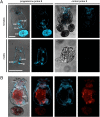
Steroids play fundamental roles regulating mammalian reproduction and development. Although sex steroids and their receptors are well characterized in vertebrates and several arthropod invertebrates, little is known about the hormones and receptors regulating reproduction in other invertebrate species. Evolutionary insights into ancient endocrine pathways can be gained by elucidating the hormones and receptors functioning in invertebrate reproduction. Using a combination of genomic analyses, receptor imaging, ligand identification, target elucidation, and exploration of function through receptor knockdown, we now show that comparable progesterone chemoreception exists in the invertebrate monogonont rotifer Brachionus manjavacas, suggesting an ancient origin of the signal transduction systems commonly associated with the development and integration of sexual behavior in mammals.
Conflict of interest statement
The authors declare no conflict of interest.
Figures


Similar articles
-
Conservation of estrogen receptor function in invertebrate reproduction.BMC Evol Biol. 2017 Mar 4;17(1):65. doi: 10.1186/s12862-017-0909-z. BMC Evol Biol. 2017. PMID: 28259146 Free PMC article.
-
Expression variations of two retinoid signaling pathway receptors in the rotifer Brachionus calyciflorus exposed to three endocrine disruptors.Ecotoxicology. 2021 Mar;30(2):343-350. doi: 10.1007/s10646-020-02339-5. Epub 2021 Jan 14. Ecotoxicology. 2021. PMID: 33443716
-
Genetic determinants of mate recognition in Brachionus manjavacas (Rotifera).BMC Biol. 2009 Sep 9;7:60. doi: 10.1186/1741-7007-7-60. BMC Biol. 2009. PMID: 19740420 Free PMC article.
-
Steroids in aquatic invertebrates.Ecotoxicology. 2007 Feb;16(1):109-30. doi: 10.1007/s10646-006-0113-1. Ecotoxicology. 2007. PMID: 17238002 Review.
-
Molecular action of progesterone.Oxf Rev Reprod Biol. 1988;10:293-347. Oxf Rev Reprod Biol. 1988. PMID: 3072504 Review. No abstract available.
Cited by
-
Genome size evolution at the speciation level: the cryptic species complex Brachionus plicatilis (Rotifera).BMC Evol Biol. 2011 Apr 7;11:90. doi: 10.1186/1471-2148-11-90. BMC Evol Biol. 2011. PMID: 21473744 Free PMC article.
-
Status in molluscan cell line development in last one decade (2010-2020): impediments and way forward.Cytotechnology. 2022 Aug;74(4):433-457. doi: 10.1007/s10616-022-00539-x. Epub 2022 Jul 24. Cytotechnology. 2022. PMID: 36110153 Free PMC article. Review.
-
Genomics of preterm birth.Cold Spring Harb Perspect Med. 2015 Feb 2;5(2):a023127. doi: 10.1101/cshperspect.a023127. Cold Spring Harb Perspect Med. 2015. PMID: 25646385 Free PMC article. Review.
-
The Genome of the Marine Rotifer Brachionus manjavacas: Genome-Wide Identification of 310 G Protein-Coupled Receptor (GPCR) Genes.Mar Biotechnol (NY). 2022 Mar;24(1):226-242. doi: 10.1007/s10126-022-10102-6. Epub 2022 Mar 9. Mar Biotechnol (NY). 2022. PMID: 35262805
-
Bromophycolide A targets heme crystallization in the human malaria parasite Plasmodium falciparum.ChemMedChem. 2011 Sep 5;6(9):1572-7. doi: 10.1002/cmdc.201100252. Epub 2011 Jul 5. ChemMedChem. 2011. PMID: 21732541 Free PMC article. No abstract available.
References
-
- Mizuta T, Kubokawa K. Presence of sex steroids and cytochrome P450 genes in amphioxus. Endocrinology. 2007;148:3554–3565. - PubMed
-
- Thornton JW, Need E, Crews D. Resurrecting the ancestral steroid receptor: ancient origin of estrogen signaling. Science. 2003;301:1714–1717. - PubMed
-
- Di Cosmo A, Di Cristo C, Paolucci M. A estradiol-17β receptor in the reproductive system of the female of Octopus vulgaris: characterization and immunolocalization. Mol Reprod Dev. 2002;61:367–375. - PubMed
Publication types
MeSH terms
Substances
LinkOut - more resources
Full Text Sources

