. 2010 Jul 6;107(27):12192-7.
doi: 10.1073/pnas.1005443107. Epub 2010 Jun 14.

Preferential localization of IgG memory B cells adjacent to contracted germinal centers

Affiliations

Preferential localization of IgG memory B cells adjacent to contracted germinal centers

Yuichi Aiba et al. Proc Natl Acad Sci U S A. .

Abstract

It has long been presumed that after leaving the germinal centers (GCs), memory B cells colonize the marginal zone or join the recirculating pool. Here we demonstrate the preferential localization of nitrophenol-chicken gamma-globulin-induced CD38(+)IgG1(+) memory B cells adjacent to contracted GCs in the spleen. The memory B cells in this region proliferated after secondary immunization, a response that was abolished by depletion of CD4(+) T cells. We also found that these IgG1(+) memory B cells could present antigen on their surface, and that this activity was required for their activation. These results implicate this peri-GC region as an important site for survival and reactivation of memory B cells.


Conflict of interest statement

The authors declare no conflict of interest.

Figures
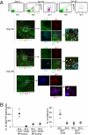
Fig. 1.
Fig. 1.
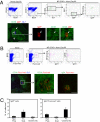
Detection of CD38+IgG1+ memory B cells adjacent to contracted GCs. (A) B6 mice were immunized with alum-precipitated NP-CGG. After 30 and 60 days, spleens were harvested and subjected to flow cytometric (Upper) and immunohistological (Lower) analyses. B220+ gated populations are shown in the upper panels. A fluorescence-activated cell sorting (FACS) profile of an unimmunized B6 mouse (day 0) is also shown. White arrows in the first and second rows of the second section indicate cells stained with both anti-CD38 and -IgG1 Abs, and in the third row indicate cells that are IgG1+λ+. [Scale bars, 150 μm (Left), 50 μm (Center).] (B) Quantification of the absolute number of memory (CD38+IgG1+) and GC (CD38IgG1+) B cells by flow cytometric (Left) and immunohistological (Right) analyses.
Fig. 2.
Fig. 2.
IgM- and IgG-type memory B cells differentially localize in the spleen. (A) B cells from B1-8hi IgH knock-in mice were adoptively transferred to CD45.1-CD45.2 F1 mice. The mice were immunized with alum-precipitated NP-CGG or left untreated. After 30 days, unimmunized mice were killed and analyzed for the presence of donor-derived, B220+CD45.1 cells by flow cytometric analysis (Upper Left; None, day 30). Immunized mice were killed at 60 days after immunization (NP-CGG + Alum, day 60). Spleens were subjected to flow cytometric (Upper) or immunohistological (Lower) analyses. [Scale bars, 300 μm (Left), 20 μm (Right).] (B) B cells purified from double-transgenic mice (B1-8hi IgH knock-in and Fucci-red) were adoptively transferred to CD45.1-CD45.2 F1 mice. The mice were immunized with alum-precipitated NP-CGG or left untreated. After 60 days, spleens from unimmunized (None) or immunized (NP-CGG + Alum, day 60) mice were subjected to flow cytometric (Upper) and immunohistological (Lower) analyses. The panels of histological analysis represent the same region in the same follicle in different sections. [Scale bars, 300 μm (Left), 50 μm (Center), 100 μm (Right).] (C) Quantification of IgMa+ cells in A and IgG1+Fucci-red+ cells in B are shown. Average number ± SD from three mice are shown. Cells in the red pulp were probably plasma cells, as judged by low expression of CD38. Fo. Scat., IgG1+ cells scattered among follicles.
Fig. 3.
Fig. 3.
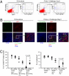
Activation of CD38+IgG1+ cells after antigen rechallenge. Fucci transgenic mice were immunized with alum-precipitated NP-CGG. After 60 days, mice were injected with NP-CGG without adjuvant (Prime + Challenged, day 2) or left untreated (Prime alone) and were killed 2 days later. Spleens were subjected to flow cytometric (A) and histological analysis (B). Percentages of CD38+Fucci and CD38+Fucci+ cells among NP-binding IgG1+ cells are indicated in the FACS profiles (A). White arrows in the right panel of B indicate the Fucci probe-positive cells stained with anti-CD38 and -IgG1 Abs. (Scale bars, 50 μm.) (C) Quantification of CD38+IgG1+ cells as either negative (FG) or positive (FG+) for the Fucci probe was performed as described in Fig. 1B. P values were calculated with a two-tailed Student's t test. P, primed alone; P+C, primed and rechallenged.
Fig. 4.
Fig. 4.
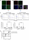
Depletion of CD4+ cells prevents activation of memory B cells and abolishes the secondary Ab response. B6 mice were immunized with alum-precipitated NP-CGG. On day 60, spleens harvested from the mice were subjected to immunohistological analysis. (A) A section stained with anti-CD38, -IgG1, and -CD4 mAbs. (Scale bar, 50 μm.) (B) A section stained with anti-PD-1, -IgG1, and -CD4 Abs. White arrows in B indicate cells stained with both anti-CD4 and -PD-1 mAbs. (Scale bar, 50 μm.) Fucci-green transgenic (C) or B6 (D) mice were immunized with alum-precipitated NP-CGG. After 60 days, mice were treated daily with anti-CD4 mAb or control rat Ab before rechallenge. Three days after the first Ab treatment, mice were rechallenged with NP-CGG without adjuvant (Prime + Challenge) or left untreated (Prime alone). (C) Spleens from Fucci-green transgenic mice were harvested 2 days after rechallenge and subjected to flow cytometric and histological analyses. Percentages of CD38+Fucci+ cells among NP-binding IgG1+ cells are indicated in the FACS profiles. (D) Anti-NP IgG1 titers in sera from immunized B6 mice were measured by ELISA 7 days after rechallenge. P, primed alone; P+C, primed and rechallenged. P values were calculated with a two-tailed Student's t test.
Fig. 5.
Fig. 5.
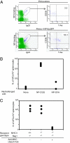
Cognate interaction with antigen-specific T cells is required for the response of IgG-type memory B cells. (A) B6 mice were immunized with alum-precipitated NP-CGG. After 60 days, mice were injected i.p. with NP-EαGFP (Prime + NP-EαGFP) or left untreated (Prime alone). After 24 h, splenocytes were analyzed by flow cytometry with Y-Ae mAb for estimating antigen presentation in the context of MHC class II. Percentages of CD38+Y-Ae+ cells among NP-binding IgG1+ cells are indicated in FACS profiles. (B and C) Memory B cells and CGG-primed T cells were purified from B6 mice immunized with alum-precipitated NP-CGG. (B) Cells were transferred to Rag1−/− mice. One day later, the mice were left untreated (None) or rechallenged with NP-CGG or NP-OVA without adjuvant. (C) Cells were transferred to Rag−/− or Rag−/− MHC class II−/− mice. After 1 day, mice were immunized with NP-CGG without adjuvant. On day 0 and day 3 after cell transfer, some mice were injected with 200 μg of Fab fragments of anti-MHC class II mAb. In both B and C, splenocytes were prepared and subjected to enzyme-linked immunospot (ELISPOT) assays for measuring the number of antibody-forming cells (AFCs) 7 days after cell transfer.

Comment in

Similar articles

Cited by

References

    1. Catron DM, Itano AA, Pape KA, Mueller DL, Jenkins MK. Visualizing the first 50 hr of the primary immune response to a soluble antigen. Immunity. 2004;21:341–347. - PubMed
    1. Allen CD, Okada T, Cyster JG. Germinal-center organization and cellular dynamics. Immunity. 2007;27:190–202. - PMC - PubMed
    1. Garside P, et al. Visualization of specific B and T lymphocyte interactions in the lymph node. Science. 1998;281:96–99. - PubMed
    1. Okada T, et al. Antigen-engaged B cells undergo chemotaxis toward the T zone and form motile conjugates with helper T cells. PLoS Biol. 2005;3:e150. - PMC - PubMed
    1. Allen CD, Okada T, Tang HL, Cyster JG. Imaging of germinal center selection events during affinity maturation. Science. 2007;315:528–531. - PubMed

Publication types

MeSH terms