Comparative Study
. 2010 Jun 29;107(26):11906-11.
doi: 10.1073/pnas.1002569107. Epub 2010 Jun 14.

Improved IL-2 immunotherapy by selective stimulation of IL-2 receptors on lymphocytes and endothelial cells

Affiliations
Comparative Study

Improved IL-2 immunotherapy by selective stimulation of IL-2 receptors on lymphocytes and endothelial cells

Carsten Krieg et al. Proc Natl Acad Sci U S A. .

Erratum in

  • Proc Natl Acad Sci U S A. 2012 Jan 3;109(1):345

Abstract

IL-2 immunotherapy is an attractive treatment option for certain metastatic cancers. However, administration of IL-2 to patients can lead, by ill-defined mechanisms, to toxic adverse effects including severe pulmonary edema. Here, we show that IL-2-induced pulmonary edema is caused by direct interaction of IL-2 with functional IL-2 receptors (IL-2R) on lung endothelial cells in vivo. Treatment of mice with high-dose IL-2 led to efficient expansion of effector immune cells expressing high levels of IL-2Rbetagamma, including CD8(+) T cells and natural killer cells, which resulted in a considerable antitumor response against s.c. and pulmonary B16 melanoma nodules. However, high-dose IL-2 treatment also affected immune cell lineage marker-negative CD31(+) pulmonary endothelial cells via binding to functional alphabetagamma IL-2Rs, expressed at low to intermediate levels on these cells, thus causing pulmonary edema. Notably, IL-2-mediated pulmonary edema was abrogated by a blocking antibody to IL-2Ralpha (CD25), genetic disruption of CD25, or the use of IL-2Rbetagamma-directed IL-2/anti-IL-2 antibody complexes, thereby interfering with IL-2 binding to IL-2Ralphabetagamma(+) pulmonary endothelial cells. Moreover, IL-2/anti-IL-2 antibody complexes led to vigorous activation of IL-2Rbetagamma(+) effector immune cells, which generated a dramatic antitumor response. Thus, IL-2/anti-IL-2 antibody complexes might improve current strategies of IL-2-based tumor immunotherapy.


Conflict of interest statement

Conflict of interest statement: O.B. is a shareholder of Nascent Biologics Inc.

Figures
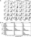
Fig. 1.
Fig. 1.
Expansion of lymphocyte subsets after stimulation with IL-2 or IL-2/mAb complexes. (A) Sorted CFSE-labeled Thy1.1+ MP CD8+ cells were transferred to WT recipients, followed by daily injections of PBS, titrated amounts of IL-2, IL-2 plus anti-IL-2 mAb MAB602 (IL-2/mAbCD122), and IL-2 plus anti-IL-2 mAb 5344 (IL-2/mAbCD25) for 5 d. On day 6, spleen cells were analyzed for CFSE profiles of donor Thy1.1+ CD8+ cells (Left) and host CD25+ FoxP3+ CD4+ Tregs (Right). (B) Total cell numbers of host CD122high MP CD8+ T cells, CD3 CD122high NK1.1+ CD49b+ NK cells, and CD25+ FoxP3+ CD4+ Tregs in spleens of animals treated as in A. Numbers to the left refer to the amount of IL-2 injected; numbers in histograms represent the percentage of divided (CFSElow) donor cells. Dashed lines refer to the levels seen in animals treated with 200,000 IU IL-2. Data are representative of four independent experiments.
Fig. 2.
Fig. 2.
Efficient control of tumor growth by IL-2/mAbCD122 complexes. (A and B) WT mice were injected s.c. with 106 B16F10 melanoma cells, followed by daily injections (indicated by gray shaded area) of PBS (Control), IL-2, IL-2/mAbCD122 complexes, or IL-2/mAbCD25 complexes. Numbers in parentheses refer to the amount of IL-2 injected. Animals were treated for 5 d starting the day after tumor inoculation (A) or for 4 d starting on day 6 after tumor inoculation (B). (C) WT mice were injected i.v. with 3 × 10⁵ B16F10 melanoma cells, followed by treatment on day 4 after injection using PBS (Control), 200,000 IU IL-2 (IL-2), 5,000 IU IL-2/mAbCD122 complexes, or 5,000 IU IL-2/mAbCD25 complexes for 5 d. Photographs of lungs are shown on day 16 after tumor inoculation. Dashed lines indicate the day maximal tumor load was reached in control mice. Data are representative of three independent experiments.
Fig. 3.
Fig. 3.
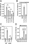
IL-2/mAbCD122 complexes show an improved profile of immune stimulation to pulmonary edema. (A) Purified CFSE-labeled Thy1.1+ MP CD8+ cells were transferred to WT recipients, followed by daily injections of PBS, graded doses of IL-2, or graded doses of IL-2/mAb complexes for 5 d. On day 6, mice were killed for determination of pulmonary wet weight (Left) and effector cell counts in host spleens (Right), including MP CD8+ (filled bars) and NK cells (open bars). Dashed lines indicate levels of pulmonary wet weight and total effector cell counts in mice receiving 200,000 IU IL-2. (B and C) WT mice were administered daily injections of PBS, 200,000 IU IL-2, 5,000 IU IL-2/mAbCD122 complexes, or 5,000 IU IL-2/mAbCD25 complexes for 5 d before staining of lungs with hematoxylin and eosin (B) or measurement of SaO2 (C). (D) Immune-depleted animals were generated by using RAG−/− mice that received sublethal irradiation (650 rad) and daily injections of depleting mAbs to Gr1 and NK1.1, followed by administration of PBS, 200,000 IU IL-2, 15,000 IU IL-2/mAbCD122 complexes, or 15,000 IU IL-2/mAbCD25 complexes before measurement of pulmonary wet weight on day 5. For B, representative regions are shown at a magnification of 40× (Upper) and 400× (Lower). Data are representative of three independent experiments.
Fig. 4.
Fig. 4.
IL-2–mediated pulmonary edema depends on CD25+ nonimmune cells. (A) WT mice received 5 daily injections of PBS or 200,000 IU IL-2 along with depleting mAbs to Thy1.2, NK1.1, CD25, or CD122. Pulmonary wet weight was determined on day 6. (B) Immune-depleted animals were generated as in Fig. 3D and received daily injections of PBS, 200,000 IU IL-2, or IL-2 plus anti-CD25 mAb. Pulmonary wet weight was determined on day 5. (C) CD25−/− mice or WT littermates received PBS or IL-2 as in A, followed by determination of pulmonary wet weight on day 6. (D) Mixed BM chimeras were generated with indicated combinations of WT or CD25−/− (KO) donor BM transferred to lethally irradiated WT or CD25−/− (KO) hosts and left for 6 wk, before treatment for 5 d with PBS or 200,000 IU IL-2. Pulmonary wet weight was calculated on day 6 by subtracting weights of PBS-treated from IL-2–treated animals. Data are representative of three independent experiments. ***P < 0.001; **P < 0.01; n.s., not significant.
Fig. 5.
Fig. 5.
Lung endothelial cells express IL-2Rs and up-regulate CD25 upon IL-2 treatment in vivo. (A) Dot plot of immune cell lineage markers (Lin) vs. CD31 showing gating on Lin+ immune cells vs. Lin CD31high endothelial cells (Upper Left) and histograms of CD25 (Upper Right), CD122 (Lower Left) and CD132 (Lower Right) expression on Lin+ immune cells (shaded area) vs. Lin CD31high endothelial cells (solid line) and isotype-matched control staining (dashed line). (B) Real-time PCR products for CD25, CD122, and CD132 of sorted WT Lin CD31+ lung cells on a 2% agarose gel are shown. GAPDH served as reference. (C) Immune-depleted mice, generated as in Fig. 3D, were left untreated or received 200,000 IU IL-2 for 4 d. On day 5, CD31+ lung endothelial cells were purified by cell sorting for Lin CD31+ cells and analyzed for CD25 mRNA expression by quantitative real-time PCR. CD25 mRNA expression is shown in arbitrary units with one SD. Data are representative of three independent experiments.
Fig. 6.
Fig. 6.
Presence of functional IL-2Rs on lung endothelial cells. (A) WT mice received a single i.v. injection of either PBS or 200,000 IU IL-2. Fifteen minutes after injection, lung cells were fixed and stained for pSTAT5 in Lin CD31+ cells. Shown are differences (Δ) in mean fluorescence intensity (MFI) of pSTAT5 staining after subtraction of isotype control stains in Lin CD31+ lung cells. (B) CD11b+ cells or Lin CD31+ cells were purified from WT lungs by using a cell sorter. Subsequently, nitrite (NO2) production in 16-h cultures was determined by using either Lin CD31+ lung cells (Right) that were left untreated (unstim.) or stimulated with IL-2 (stim.). Unstimulated (unstim.) or IFN-γ/LPS-stimulated (stim.) CD11b+ cells (Left) served as controls. The detection limit, indicated as dashed line, was at 6 μM. Data are representative of two independent experiments. **P < 0.01; ***P < 0.001.

References

    1. Malek TR. The biology of interleukin-2. Annu Rev Immunol. 2008;26:453–479. - PubMed
    1. Boyman O, Purton JF, Surh CD, Sprent J. Cytokines and T-cell homeostasis. Curr Opin Immunol. 2007;19:320–326. - PubMed
    1. Minami Y, Kono T, Miyazaki T, Taniguchi T. The IL-2 receptor complex: Its structure, function, and target genes. Annu Rev Immunol. 1993;11:245–268. - PubMed
    1. Sakaguchi S, Sakaguchi N, Asano M, Itoh M, Toda M. Immunologic self-tolerance maintained by activated T cells expressing IL-2 receptor alpha-chains (CD25). Breakdown of a single mechanism of self-tolerance causes various autoimmune diseases. J Immunol. 1995;155:1151–1164. - PubMed
    1. Lefrancois L, Klein JR, Paetkau V, Bevan MJ. Antigen-independent activation of memory cytotoxic T cells by interleukin 2. J Immunol. 1984;132:1845–1850. - PubMed

Publication types

MeSH terms

LinkOut - more resources