Review
. 2010 Jun 14;189(6):925-35.
doi: 10.1083/jcb.201002021.

Regulation of innate immune responses by autophagy-related proteins

Affiliations
Review

Regulation of innate immune responses by autophagy-related proteins

Tatsuya Saitoh et al. J Cell Biol. .

Abstract

Pattern recognition receptors detect microbial components and induce innate immune responses, the first line of host defense against infectious agents. However, aberrant activation of immune responses often causes massive inflammation, leading to the development of autoimmune diseases. Therefore, both activation and inactivation of innate immune responses must be strictly controlled. Recent studies have shown that the cellular machinery associated with protein degradation, such as autophagy, is important for the regulation of innate immunity. These studies reveal that autophagy-related proteins are involved in the innate immune response and may contribute to the development of inflammatory disorders.


Figures

Figure 1.
Figure 1.
Atgs are essential components for autophagosome formation. Autophagy, a bulk degradation system, is induced after sensing various types of stress, such as nutrient starvation, the accumulation of protein aggregates, and invasion by microbes. This system enables the reuse of cellular components and the clearance of unfavorable substances, thus contributing to the maintenance of cellular homeostasis and the prevention of disease. Atgs are recruited to the isolation membrane, a source membrane of autophagosomes, and play a vital role in driving the membrane trafficking necessary for the generation of autophagosomes. Autophagosomes then fuse with lysosomes to become autolysosomes, leading to the degradation of the constituents trapped inside. PE, phosphatidylethanolamine.
Figure 2.
Figure 2.
TLR response and Atgs. (A) Atgs control endotoxin-induced IL-1β production. TLR4 triggers both MyD88- and TRIF-dependent signaling pathways after sensing LPS. The IKK-α–IKK-β–NEMO complex mediates the activation of the transcription factor NF-κB, which in turn induces the transcription of proinflammatory cytokines and pro–IL-1β. The TBK1–IKK-i complex mediates the activation of the transcription factor IRF3, which then induces the transcription of type I IFNs and IFN-inducible genes. In autophagy-deficient cells, high levels of ROS are generated, which mediate TRIF-dependent caspase-1 activation, resulting in the processing of IL-1β. However, in wild-type macrophages, limited amounts of IL-1β are induced by LPS as the result of a lack of ROS generation. (B) Atgs contribute to TLR-dependent elimination of pathogens. After detection of the fungal cell wall component zymosan, TLR2 induces the maturation of phagosomes, leading to the elimination of the fungus. Atgs such as Atg5, Atg7, and PI3K are involved in the fusion of phagosomes with lysosomes. (C) Ligands for TLR7 such as ssRNA and imiquimod induce the formation of autophagosomes via MyD88, an essential adaptor molecule, and promote the elimination of Bacillus Calmette-Guerin. LPS, a ligand for TLR4, induces the formation of autophagosomes via the TRIF-p38 signaling axis, leading to the elimination of Mycobacterial bacilli. Atgs such as Atg5, beclin, and PI3K are required for the formation of autophagosomes by TLR stimulation.
Figure 3.
Figure 3.
Sensing of bacterial polypeptides by Nod1 and Nod2 triggers the formation of autophagosomes. Nod1 and Nod2 are composed of N-terminal CARDs, a centrally located nucleotide-binding oligomerization domain (NOD), and multiple C-terminal leucine-rich repeats (LRRs). The leucine-rich repeats of Nod1 and Nod2 detect iE-DAP and MDP, respectively. After sensing these ligands, Nod1 and Nod2 induce the formation of autophagosomes, leading to the promotion of antigen presentation and the enhancement of bactericidal responses. Atgs such as Atg5, Atg7, and Atg16L1 are involved in Nod1- and Nod2-mediated formation of autophagosomes.
Figure 4.
Figure 4.
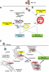
Regulation of nucleic acid–induced type I IFN production by Atgs. (A) RIG-I and MDA-5, CARD-containing RNA helicases, sense viral dsRNA and signal to IPS-1, a CARD-containing adaptor molecule, to induce the expression of type I IFN/IFN-inducible genes and proinflammatory cytokines. Atg12-Atg5 conjugates are recruited to the CARD domains of RIG-I, MDA-5, and IPS-1 and suppress the dsRNA-induced innate immune response. (B) TLR7 detects viral ssRNA and induces innate immune responses. Targeted disruption of Atg5 or pharmacological inhibition of PI3K results in the impairment of TLR7-mediated expression of type I IFN/IFN-inducible genes and proinflammatory cytokines. (C) STING, a multispanning membrane protein, and TBK1, an IRF3 kinase, mediate the induction of innate immune responses by dsDNA. After sensing dsDNA, STING moves from the ER to the Golgi apparatus and finally reaches the cytoplasmic punctate structures to assemble with TBK1, resulting in the IRF3-dependent expression of type I IFNs/IFN-inducible genes. STING colocalizes with Atg9a in the Golgi apparatus after dsDNA stimulation. The loss of Atg9a greatly enhances the dsDNA-induced assembly of STING and TBK1, leading to aberrant activation of the innate immune response.
Figure 5.
Figure 5.
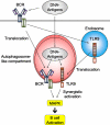
DNA-containing antigens induce TLR9 signal from autophagosome-like compartments. DNA-containing antigens stimulate BCRs, leading to the translocation of TLR9 from the endosomes to autophagosome-like compartments. DNA-containing antigens also promote the movement of BCRs from the cell surface to autophagosome-like compartments. In these autophagosome-like compartments, DNA-containing antigens trigger both BCR and TLR9 signaling pathways, resulting in the synergistic activation of MAPK. Functional PLD and microtubules are required for the translocation of these receptors and play a critical role in the hyperresponse to DNA-containing antigens.

References

    1. Berón W., Gutierrez M.G., Rabinovitch M., Colombo M.I. 2002. Coxiella burnetii localizes in a Rab7-labeled compartment with autophagic characteristics. Infect. Immun. 70:5816–5821 10.1128/IAI.70.10.5816-5821.2002 - DOI - PMC - PubMed
    1. Beutler B. 2009. Microbe sensing, positive feedback loops, and the pathogenesis of inflammatory diseases. Immunol. Rev. 227:248–263 10.1111/j.1600-065X.2008.00733.x - DOI - PMC - PubMed
    1. Bhoj V.G., Chen Z.J. 2009. Ubiquitylation in innate and adaptive immunity. Nature. 458:430–437 10.1038/nature07959 - DOI - PubMed
    1. Bjørkøy G., Lamark T., Brech A., Outzen H., Perander M., Overvatn A., Stenmark H., Johansen T. 2005. p62/SQSTM1 forms protein aggregates degraded by autophagy and has a protective effect on huntingtin-induced cell death. J. Cell Biol. 171:603–614 10.1083/jcb.200507002 - DOI - PMC - PubMed
    1. Blander J.M., Medzhitov R. 2004. Regulation of phagosome maturation by signals from toll-like receptors. Science. 304:1014–1018 10.1126/science.1096158 - DOI - PubMed

MeSH terms