. 2010 Jun 10;5(6):e11045.
doi: 10.1371/journal.pone.0011045.

Quercetin targets cysteine string protein (CSPalpha) and impairs synaptic transmission

Affiliations

Quercetin targets cysteine string protein (CSPalpha) and impairs synaptic transmission

Fenglian Xu et al. PLoS One. .

Abstract

Background: Cysteine string protein (CSPalpha) is a synaptic vesicle protein that displays unique anti-neurodegenerative properties. CSPalpha is a member of the conserved J protein family, also called the Hsp40 (heat shock protein of 40 kDa) protein family, whose importance in protein folding has been recognized for many years. Deletion of the CSPalpha in mice results in knockout mice that are normal for the first 2-3 weeks of life followed by an unexplained presynaptic neurodegeneration and premature death. How CSPalpha prevents neurodegeneration is currently not known. As a neuroprotective synaptic vesicle protein, CSPalpha represents a promising therapeutic target for the prevention of neurodegenerative disorders.

Methodology/principal findings: Here, we demonstrate that the flavonoid quercetin promotes formation of stable CSPalpha-CSPalpha dimers and that quercetin-induced dimerization is dependent on the unique cysteine string region. Furthermore, in primary cultures of Lymnaea neurons, quercetin induction of CSPalpha dimers correlates with an inhibition of synapse formation and synaptic transmission suggesting that quercetin interfers with CSPalpha function. Quercetin's action on CSPalpha is concentration dependent and does not promote dimerization of other synaptic proteins or other J protein family members and reduces the assembly of CSPalpha:Hsc70 units (70kDa heat shock cognate protein).

Conclusions/significance: Quercetin is a plant derived flavonoid and popular nutritional supplement proposed to prevent memory loss and altitude sickness among other ailments, although its precise mechanism(s) of action has been unclear. In view of the therapeutic promise of upregulation of CSPalpha and the undesired consequences of CSPalpha dysfunction, our data establish an essential proof of principle that pharmaceutical agents can selectively target the neuroprotective J protein CSPalpha.


Conflict of interest statement

Competing Interests: The authors have declared that no competing interests exist.

Figures

Figure 1
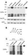
Figure 1. Quercetin promotes CSPα dimerization.
(A) CAD cells were transiently transfected with 0.5 µg of c-myc-CSPα DNA and treated with the indicated agent (200 µM quercetin, 1 µM geldanamycin, 1.5 µM MPP+, 0.2 mM H2O2 or 10 µM lactacystin) for 24 hours prior to lysis. 40 µg of cellular protein was resolved by SDS-PAGE and CSPα and Hsc70 were detected by Western analysis. β-actin is shown as a loading control. (B) Chemical structure of quercetin dihydrate. (C) CAD cells were transfected with 1.0 µg of c-myc-CSPα DNA and treated with 100 µM quercetin for 24 hours prior to lysis. 30 µg of protein was heated at either 37°C or 80°C for 10 minutes prior to being resolved on an SDS-PAGE gel. CSPα was detected by Western analysis with a c-myc antibody. Arrows indicate CSPα dimer at ∼72 kDa; asterisks indicates palmitoylated CSPα monomer at ∼34 kDa. Data are representative of three separate experiments.
Figure 2
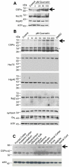
Figure 2. Quercetin exerts a concentration-dependent effect on the formation of the CSPα dimer in rat cortical neurons and CAD cells.
(A) Western blot of cultured rat cortical neurons were treated with indicated concentrations of quercetin for 24 hours prior to lysis. Equal amounts of cellular protein were resolved by SDS-PAGE as confirmed by ponceau S staining. (B) CAD cells were transiently transfected with 1.0 µg c-myc-CSPα DNA and treated with indicated concentrations of quercetin for 24 hours prior to lysis. Following separation of cellular protein (30 µg) by SDS-PAGE, CSPα, Hsc70, Hsp40, Rdj2, syntaxin and Gαs were detected by Western analysis. β-actin is shown as a loading control. Data are representative of three separate experiments. (C) Native CSPα was detected in adult rat brain by Western analysis with a monoclonal anti-CSPα antibody. Twenty-five micrograms of unfractionated tissue homogenate isolated from the indicated regions of rat brain were separated by SDS-PAGE, transferred to PVDF and probed. Arrows indicate the CSPα dimer at ∼72 kDa; * indicates a palmitoylated CSPα monomer at ∼34 kDa. Actin is shown as a loading control.
Figure 3
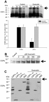
Figure 3. Quercetin increases the stability of the CSPα dimer in CAD cells.
(A) CAD cells were transfected with 1.0 µg of c-myc CSPα DNA and treated with 100 µM quercetin as shown. Cells were lysed at indicated times following quercetin treatment. 30 µg of protein was resolved. Upper panel: CSPα was detected by Western analysis with the c-myc antibody. Lower panel: Quantification of the CSPα dimer at 5 minutes and 24 hours in control and quercetin-treated cells. Results are expressed as mean +/− SE for a total of 4 separate experiments. (B) Equal volumes of purified recombinant rat CSPα were treated with indicated concentrations of quercetin for 24 hrs (C) CAD cells were transfected with 1.0 µg CSPα1–198, CSPα1–82, CSPαΔc and Rdj2 DNA and treated with 100 µM quercetin for 24 hours. Arrows indicate CSPα dimer at ∼72 kDa. Data are representative of three separate experiments.
Figure 4
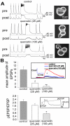
Figure 4. Quercetin inhibits synapse formation in Lymnaea stagnalis primary cultured neurons.
The presynaptic, cholinergic neuron, visceral dorsal 4 (VD4) and the postsynaptic neuron, left pedal dorsal 1 (LPeD1) were juxtaposed and cultured in the absence or presence of quercetin (25 or 100 µM) for 12–18 hours. Prior to intracellular recordings, quercetin was washed off. (A) Sample traces of presynaptic action potentials on VD4 cells and excitatory postsynaptic potentials (EPSPs) on LPeD1 cells. (B) The mean amplitude of EPSP and the ratio of potentiated EPSP (pEPSP) over EPSP was reduced in the presence of both 25 µM quercetin (n = 5) and 100 µM quercetin (n = 7) (inserts). Statistical significance was determined using Students' t-test. * indicates significant difference at the level of p = 0.05. Error bars indicate S.E.
Figure 5
Figure 5. Quercetin induces the CSPα dimer in Lymnaea stagnalis.
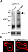
(A) Lymnaea were maintained in pond water containing 100 µM quercetin over night as indicated prior to harvesting of the VD4 ganglia from several snails. Equal numbers of ganglia were combined and resolved on a gel. Native CSPα was detected by Western analysis with the CSPα polyclonal antibody. The panels shown are from the same experiment and are representative of three independent experiments. Arrow indicates CSPα dimer at ∼72 kDa. Hsc70 is shown as a loading control. (B) Paired Lymnaea soma were cultured overnight and subjected to immunostaining with CSPα polyclonal antibody. Stacks of 0.28 µm slices were collected and collapsed into Z projections in maximum intensity using ImageJ. Images are representative of five experiments.
Figure 6
Figure 6. Acute application of quercetin blocks synaptic transmission in Lymnaea neurons.
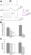
VD4 (presynaptic) and LPeD1 (postsynaptic) were co-cultured overnight, presynaptic action potentials were induced and the amplitude of the excitatory post synaptic potential (EPSPs) was measured. (A) Quercetin increased the presynaptic repolarization phase of the action potential by predominately rendering the neuron incapable of firing continuously (clamping of tetanic bursts) *. The EPSP amplitude was greatly diminished within minutes of perfusion with 100 µM quercetin. A typical action potential and EPSP are shown before (red) and after (blue) exposure to quercetin. (B) Summary of the effect of quercetin (25 and 100 µM) and following wash out (10–20 mins) on the mean amplitude of action potential-generated EPSPs. (C) Summary of the effect of quercetin (25 and 100 µM) and quercetin washout on the ratio of potentiated EPSP (LPeD1) over EPSP. Statistical significance was determined using Students' paired t-test. * indicates significant difference at the level of p = 0.05. Error bars indicate S.E.
Figure 7
Figure 7. Quercetin does not alter the postsynaptic left pedal dorsal 1 (LPeD1) response to acetylcholine, but reduces action potential or depolarization-induced Ca2+ entry into the presynaptic neuron.
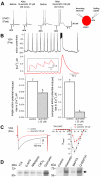
(A) LPeD1 cells were cultured. Acetylcholine (ACh, 1 µM) was exogenously applied before and after exposure to quercetin (25 or 100 µM) for 30 mins and postsynaptic potential was monitored. (B) Intracellular recording of action potentials from cultured VD4 neurons before and after perfusion of quercetin (25 µM) were simultaneously made with the measurement of cytoplasmic Ca2+ concentrations using a Ca2+ imaging technique. Single action potentials induced corresponding Ca2+ transients (insert) and burst of action potentials triggered a robust compound Ca2+ rise. However the mean values of [Ca2+] rise in response to single and bursts of action potential were significantly reduced after exposure to quercetin for 20 mins (n = 4). (C) Quercetin inhibited voltage-gated Ca2+ currents (ICa) in presynaptic VD4 neurons elicited by step depolarization of cells from −80 mV to +70 mV in 10 mV increments before and after exposure to quercetin 100 µM for 10 mins. The left panal shows representative raw traces of ICa evoked by a square depolarization pulse from −80 mV to +20 mV for 500 ms. The right panel shows normalized current-voltage relations of ICa (n = 4). (D) CAD cells were transiently transfected with 0.75 µg CSPα, treated with 1 µM tetrotodoxin (TTX), 50 µM D-2-amino-5-phosphonovalerate (D-APV), 50 µm nifedipine, 100 µm CdCl2 or 100 µM quercetin for 24 hours. Right panel: CAD cells were treated with 100 µM quercetin for 24 hour, 20 µM BAPTA-AM 1 hour as indicated and 30 µg of cell lysate was resolved by SDS-PAGE. Data are representative of three separate experiments.
Figure 8
Figure 8. Quercetin reduces CSPα:Hsc70 association.
Co-immunoprecipitation of CSPα and Hsc70 from control and quercetin-treated CAD cells followed by Western analysis. Immunoprecipitation was achieved by incubating 300 µg CAD cell lysate with anti-myc monoclonal, immunoprecipitated proteins were separated by SDS-PAGE and evaluated by Western analysis with anti-CSPα polyclonal and anti-Hsc70 monoclonal. Arrow indicates CSPα dimer at ∼72 kDa. Data are representative of three separate experiments.
Figure 9
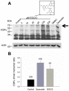
Figure 9. Epigallocatechin gallate (EGCG) stimulates formation of CSPα-CSPα dimers.
(A) CAD cells were transiently transfected with 1.0 µg c-myc-CSPα DNA and treated with indicated concentrations of EGCG for 24 hours prior to lysis. Following separation of cellular protein (30 µg) by SDS-PAGE, CSPα, was detected by Western analysis. β-actin is shown as a loading control. Arrow indicates CSPα dimer at ∼72 kDa. (B) Quantification of the CSPα dimer to monomeric palmitoylated CSPα ratio under control, 100 µM quercetin and 200 µm EGCG conditions. Numbers in parentheses indicate the numbers of experiments; error bars denote standard errors.
Figure 10
Figure 10. Model depicting the inhibition of CSPα chaperone activity by quercetin.
The synaptic vesicle protein CSPα has unique anti-neurodegenerative properties. Distinct CSPα complexes exist: inactive, active (in complex with Hsc70 and SGT), and a CSPα dimer. Quercetin promotes the CSPα dimer, inhibits assembly of the active CSPα complex and synaptic transmission.

Similar articles

Cited by

References

    1. Zhao X, Braun AP, Braun JE. Biological Roles of Neural J Proteins. Cell Mol Life Sci. 2008;65:2385–2396. - PMC - PubMed
    1. Mastrogiacomo A, Parsons SM, Zampighi GA, Jenden DJ, Umbach JA, et al. Cysteine string proteins: a potential link between synaptic vesicles and presynaptic Ca2+ channels. Science. 1994;263:981–982. - PubMed
    1. Blondeau F, Ritter B, Allaire PD, Wasiak S, Girard M, et al. Tandem MS analysis of brain clathrin-coated vesicles reveals their critical involvement in synaptic vesicle recycling. Proc Natl Acad Sci U S A. 2004;101:3833–3838. - PMC - PubMed
    1. Zinsmaier KE, Eberle KK, Buchner E, Walter N, Benzer S. Paralysis and early death in cysteine string protein mutants of Drosophila. Science. 1994;263:977–980. - PubMed
    1. Fernandez-Chacon R, Wolfel M, Nishimune H, Tabares L, Schmitz F, et al. The synaptic vesicle protein CSP alpha prevents presynaptic degeneration. Neuron. 2004;42:237–251. - PubMed

Publication types