

The Investigation on the Distribution of Malassezia Yeasts on the Normal Korean Skin by 26S rDNA PCR-RFLP
- PMID: 20548850
- PMCID: PMC2883363
- DOI: 10.5021/ad.2009.21.1.18
The Investigation on the Distribution of Malassezia Yeasts on the Normal Korean Skin by 26S rDNA PCR-RFLP
Abstract
Background: Malassezia yeasts are normal flora of the skin that are discovered in 75~98% of health subjects, but since its association with various skin disorders have been known, many studies have been conducted in the distribution of the yeasts.
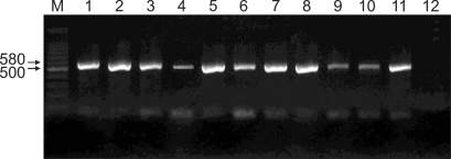
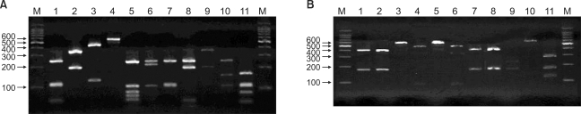
Objective: To isolate, identify, and classify Malassezia yeasts from the normal human skin of Koreans by using the rapid and accurate molecular biology method (26S rDNA PCR-RFLP) which overcome the limits of morphological and biochemical methods, and to gather a basic database that will show its relation to various skin diseases.
Methods: Malassezia yeasts were cultured from clinically healthy human skin using scrub-wash technique at five sites (forehead, cheek, chest, upper arm, and thigh) and swabbing technique at scalp in 160 participants comprised of 80 males and 80 females aged from 0 to 80. Identification of obtained strains were placed into the one of the 11 species by 26S rDNA PCR-RFLP.
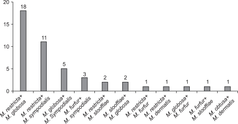
Results: An overall positive culture rate was 62.4% (599/960). As shown in the experiment groups by their age, the positive culture rate was the highest (74.2%) in the age 21~30 and 31~40 (89/120). In the experiment groups by different body areas, the scalp showed the highest positive culture rate of 90% (144/160). On analysis of 26S rDNA PCR-RFLP, M. globosa was the most predominant species in the age 0~10 (32.8%), 11~20 (28.9%), 21~30 (32.3%). M. restricta was identified as predominant species in the age 41~50 (27.9%), 61~70 (31.5%) and 71~80 (24.0%). In the age 31~40 years, M. sympodialis was found to be the most common species (24.6%). According to body site, M. restricta was more frequently recovered in the scalp (56.8%), forehead (39.8%) and cheek (24.0%) and while M. globosa was more frequently recovered in the chest (36.8%). Higher positive culture rates of Malassezia yeasts were shown in male subjects than female counterparts in all body areas except scalp (p<0.05). Especially in this study, M. dermatis, newly isolated Malassezia species from atopic dermatitis patient in Japan, was isolated and identified in 19 cases (1.9%) in healthy subjects.
Conclusion: The key is to recognize the existence of a difference in the type of Malassezia species in different ages as well as body areas, which reflects differing skin lipid levels in various ages and different body areas. Moreover, 26S rDNA PCR-RFLP analysis which was opted in this study could provide a sensitive and rapid identification system for Malassezia species, which may be applied to epidemiological surveys and clinical practice.
Keywords: 26S rDNA PCR-RFLP; Malassezia.
Figures
Similar articles
-
Molecular analysis of malassezia microflora on the skin of the patients with atopic dermatitis.Ann Dermatol. 2010 Feb;22(1):41-7. doi: 10.5021/ad.2010.22.1.41. Epub 2010 Feb 28. Ann Dermatol. 2010. PMID: 20548879 Free PMC article.
-
Epidemiologic Study of Malassezia Yeasts in Patients with Malassezia Folliculitis by 26S rDNA PCR-RFLP Analysis.Ann Dermatol. 2011 May;23(2):177-84. doi: 10.5021/ad.2011.23.2.177. Epub 2011 May 27. Ann Dermatol. 2011. PMID: 21747616 Free PMC article.
-
Epidemiologic Study of Malassezia Yeasts in Seborrheic Dermatitis Patients by the Analysis of 26S rDNA PCR-RFLP.Ann Dermatol. 2010 May;22(2):149-55. doi: 10.5021/ad.2010.22.2.149. Epub 2010 May 17. Ann Dermatol. 2010. PMID: 20548904 Free PMC article.
-
Malassezia species in healthy skin and in dermatological conditions.Int J Dermatol. 2016 May;55(5):494-504. doi: 10.1111/ijd.13116. Epub 2015 Dec 29. Int J Dermatol. 2016. PMID: 26710919 Review.
-
[A study of culture-based easy identification system for Malassezia].Med Mycol J. 2011;52(4):297-303. doi: 10.3314/mmj.52.297. Med Mycol J. 2011. PMID: 22123328 Review. Japanese.
Cited by
-
Distribution of malassezia species on the scalp in korean seborrheic dermatitis patients.Ann Dermatol. 2011 May;23(2):156-61. doi: 10.5021/ad.2011.23.2.156. Epub 2011 May 27. Ann Dermatol. 2011. PMID: 21747613 Free PMC article.
-
Malassezia spp. Yeasts of Emerging Concern in Fungemia.Front Cell Infect Microbiol. 2020 Jul 28;10:370. doi: 10.3389/fcimb.2020.00370. eCollection 2020. Front Cell Infect Microbiol. 2020. PMID: 32850475 Free PMC article. Review.
-
Putting It All Together to Understand the Role of Malassezia spp. in Dandruff Etiology.Mycopathologia. 2018 Dec;183(6):893-903. doi: 10.1007/s11046-018-0283-4. Epub 2018 Jun 26. Mycopathologia. 2018. PMID: 29946996 Review.
-
The itchy scalp--scratching for an explanation.Exp Dermatol. 2011 Dec;20(12):959-68. doi: 10.1111/j.1600-0625.2011.01389.x. Exp Dermatol. 2011. PMID: 22092575 Free PMC article. Review.
-
Molecular analysis of malassezia microflora on the skin of the patients with atopic dermatitis.Ann Dermatol. 2010 Feb;22(1):41-7. doi: 10.5021/ad.2010.22.1.41. Epub 2010 Feb 28. Ann Dermatol. 2010. PMID: 20548879 Free PMC article.
References
-
- Janik MP, Heffernan MP. Yeasts infection: Candidiasis, Pityriasis (Tinea) versicolor. In: Wolff K, Goldsmith LA, Katz SI, Gilchrest BA, Paller AS, Leffell DJ, editors. Fitzpatrick's dermatology in general medicine. 7th ed. New York: McGraw-Hill; 2008. pp. 1822–1830.
-
- Ahn KJ. Taxonomy of the genus Malassezia. Korean J Med Mycol. 1998;3:81–88.
-
- Ljubojević S, Skerlev M, Lipozencić J, Basta-Juzbasic A. The role of Malassezia furfur in dermatology. Clin Dermatol. 2002;20:179–182. - PubMed
-
- Kanda N, Tani K, Enomoto U, Nakai K, Watanabe S. The skin fungus-induced Th1- and Th2-related cytokine, chemokine and prostaglandin E2 production in peripheral blood mononuclear cells from patients with atopic dermatitis and psoriasis vulgaris. Clin Exp Allergy. 2002;32:1243–1250. - PubMed
-
- Ginarte M, Fabeiro JM, Toribio J. Confluent and reticulated papillomatosis (Gougerot-Carteaud) successfully treated with tacalcitol. J Dermatolog Treat. 2002;13:27–30. - PubMed
LinkOut - more resources
Full Text Sources
Research Materials
