

Aneurysmal benign fibrous histiocytoma with atrophic features
- PMID: 20548854
- PMCID: PMC2883367
- DOI: 10.5021/ad.2009.21.1.42
Aneurysmal benign fibrous histiocytoma with atrophic features
Abstract
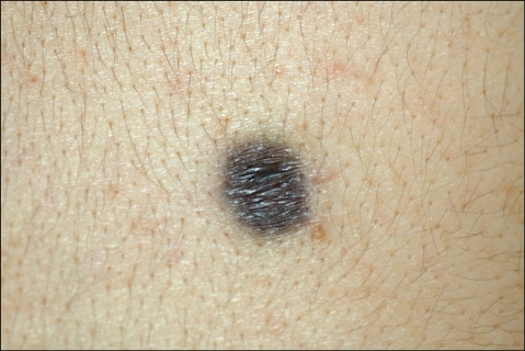
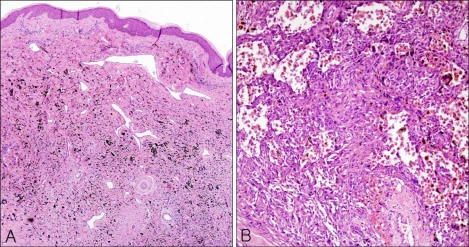
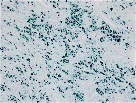
Aneurysmal benign fibrous histiocytoma is an uncommon pathologic variant of dermatofibroma. In addition to the features of a typical dermatofibroma, it has large cleft-like or cavernous blood-filled spaces with numerous hemosiderin pigments. It should be differentiated from angiomatoid malignant fibrous histiocytoma, malignant melanoma, and vascular tumors such as Kaposi's sarcoma and angiosarcoma. Atrophic dermatofibroma is also a rare variant of dermatofibroma, and the combination of aneurysmal and atrophic features is rarer still. We report a case of aneurysmal benign fibrous histiocytoma with atrophic features in a 27-year-old male who had a grayish-brown atrophic patchy lesion on his back for 2 years.
Keywords: Aneurysmal variant; Atrophy; Dermatofibroma.
Figures
References
-
- Santa Cruz DJ, Kyriakos M. Aneurysmal ("angiomatoid") fibrous histiocytoma of the skin. Cancer. 1981;47:2053–2061. - PubMed
-
- Calonje E, Fletcher CD. Aneurysmal benign fibrous histiocytoma: clinicopathological analysis of 40 cases of a tumour frequently misdiagnosed as a vascular neoplasm. Histopathology. 1995;26:323–331. - PubMed
-
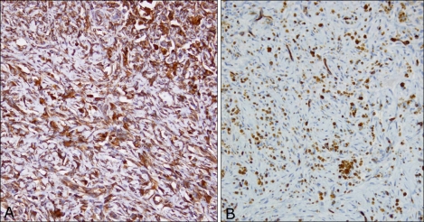
- Goldblum JR, Tuthill RJ. CD34 and factor-XIIIa immunoreactivity in dermatofibrosarcoma protuberans and dermatofibroma. Am J Dermatopathol. 1997;19:147–153. - PubMed
-
- Page EH, Assaad DM. Atrophic dermatofibroma and dermatofibrosarcoma protuberans. J Am Acad Dermatol. 1987;17:947–950. - PubMed
Publication types
LinkOut - more resources
Full Text Sources
