

A case of isolated plexiform neurofibroma in a patient with myasthenia gravis
- PMID: 20548857
- PMCID: PMC2883370
- DOI: 10.5021/ad.2009.21.1.53
A case of isolated plexiform neurofibroma in a patient with myasthenia gravis
Abstract
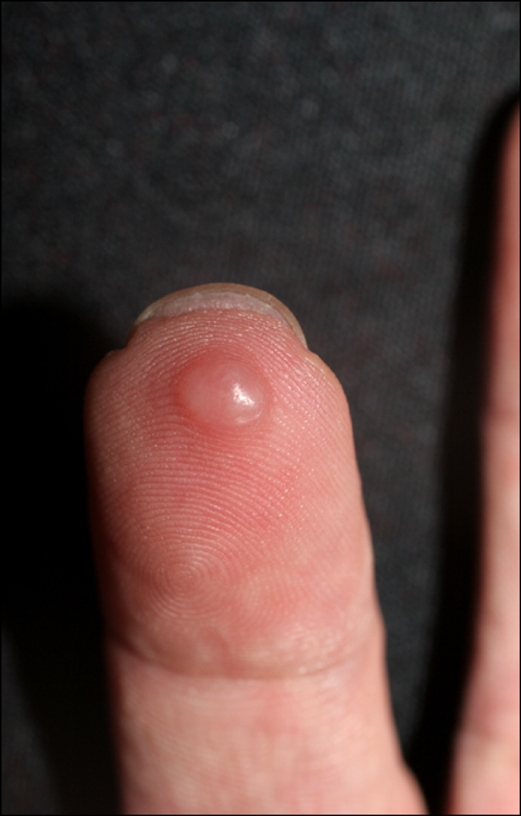
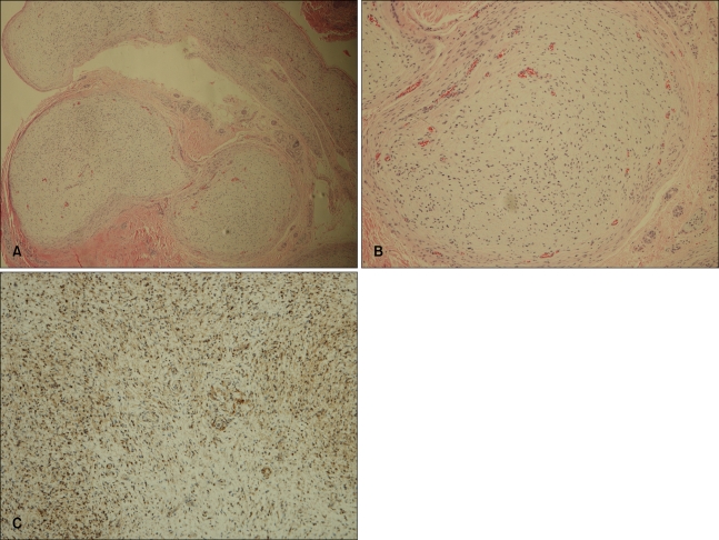
We report a case of an isolated plexiform neurofibroma occurring in a patient with myasthenia gravis. A 48-year-old man presented with asymptomatic skin-colored nodules on the tip of his 4th finger. Microscopically, a plexiform neurofibroma was identified located in the dermis that appeared to originate from small superficial nerves. He had a 20-year history of treated myasthenia gravis; otherwise, his personal and family histories were unremarkable. Given that myasthenia gravis is a disorder of the peripheral nerves, plexiform neurofibromas could be associated with myasthenia gravis. However, the development of an isolated plexiform neurofibroma in a case of myasthenia gravis has not yet been reported. The occurrence of a neurofibromas in a patient with myasthenia gravis suggests a link in the pathogenesis of these two diseases.
Keywords: Isolated; Myasthenia gravis; Plexiform neurofibroma.
Figures
References
-
- Robert L, Joel C. The neurofibromatoses. In: Wolff K, Goldsmith LA, Katz SI, Gilchrest BA, Paller AS, Leffell DJ, editors. Fitzpatrick's dermatology in general medicine. 7th ed. New York: McGraw-Hill; 2008. pp. 1332–1334.
-
- Legius E, Descheemaeker MJ, Fryns JP, Van den Berghe H. Neurofibromatosis type 1. Genet Couns. 1994;5:225–241. - PubMed
-
- Kubota A, Komiyama A, Hasegawa O. Myasthenia gravis and alopecia areata. Neurology. 1997;48:774–775. - PubMed
-
- Younus J, Ahmed AR. The relationship of pemphigus to neoplasia. J Am Acad Dermatol. 1990;23:498–502. - PubMed
-
- Kwan SY, Lin JH, Su MS. Coexistence of epilepsy, myasthenia gravis and psoriasis vulgaris. Zhonghua Yi Xue Za Zhi (Taipei) 2000;63:153–157. - PubMed
Publication types
LinkOut - more resources
Full Text Sources
