

Case Reports
doi: 10.5021/ad.2009.21.1.75.
Epub 2009 Feb 28.
A Case of Vitiligo after Kawasaki's Disease
Affiliations
- PMID: 20548863
- PMCID: PMC2883377
- DOI: 10.5021/ad.2009.21.1.75
Item in Clipboard
Case Reports
A Case of Vitiligo after Kawasaki's Disease
Ann Dermatol.
2009 Feb.
Abstract
Vitiligo is a common skin disease, but its pathogenesis has not been fully determined, though an autoimmune etiology is considered likely. Kawasaki disease (KD) is an acute multisystem vasculitis of childhood associated with coronary arteriopathy, and is diagnosed based on clinical criteria. Furthermore, vitiligo has been associated with several other diseases, but no report has been issued about the relationship between vitiligo and Kawasaki's disease. The author's report the case of an 8-year-old male child that presented with depigmented lesions, which developed from the desquamative skin lesions of Kawasaki's disease.
Keywords: Kawasaki's disease; Vitiligo.
Figures
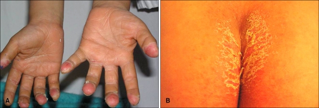
Characteristic erythematous, desquamative maculopatches on the fingertips (A) and perianal skin (B) on Feb. 10, 2007, when he was admitted to our hospital.
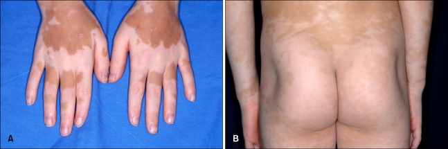
At four months after KD onset, depigmented maculopatches were noted on the dorsa of both hands (A), forearms, and buttocks (B). Previously, these sites had been affected by typical KD skin rashes.
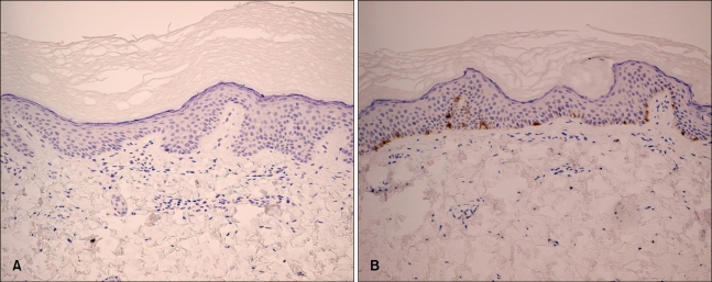
Histological examinations of a hypopigmented patch revealed the absence of epidermal melanocytes, which was confirmed by immunostaining for melan A (×200) (A). Perilesional areas showed normal epidermal melanocyte levels (×200) (B).
Similar articles
-
Adult Kawasaki's disease with myocarditis, splenomegaly, and highly elevated serum ferritin levels.Heart Lung. 2010 Mar-Apr;39(2):164-72. doi: 10.1016/j.hrtlng.2009.06.007. Epub 2009 Sep 3. Heart Lung. 2010. PMID: 20207278 Free PMC article.
-
[Adult multiple coronary aneurysms of Kawasaki's disease's sequelae; two autopsy cases].Rinsho Byori. 1998 Feb;46(2):177-81. Rinsho Byori. 1998. PMID: 9528343 Japanese.
-
Kawasaki's disease, acrodynia, and mercury.Curr Med Chem. 2008;15(28):3000-10. doi: 10.2174/092986708786848712. Curr Med Chem. 2008. PMID: 19075648 Review.
-
Kawasaki's disease: An unusual presentation.J Cardiovasc Dis Res. 2012 Jul;3(3):240-1. doi: 10.4103/0975-3583.98902. J Cardiovasc Dis Res. 2012. PMID: 22923945 Free PMC article.
-
Pathogenesis and treatment of Kawasaki's disease.Curr Opin Rheumatol. 2005 Sep;17(5):617-23. doi: 10.1097/01.bor.0000174184.15901.ee. Curr Opin Rheumatol. 2005. PMID: 16093842 Review.
Cited by
-
Tetrahydrobiopterin as a Trigger for Vitiligo: Phototransformation during UV Irradiation.Int J Mol Sci. 2023 Sep 1;24(17):13586. doi: 10.3390/ijms241713586. Int J Mol Sci. 2023. PMID: 37686391 Free PMC article.
-
How Should We Classify Kawasaki Disease?Front Immunol. 2018 Dec 14;9:2974. doi: 10.3389/fimmu.2018.02974. eCollection 2018. Front Immunol. 2018. PMID: 30619331 Free PMC article. Review.
References
-
- Kovacs SO. Vitiligo. J Am Acad Dermatol. 1998;38:647–666. - PubMed
-
- Kemp EH, Gavalas NG, Gawkrodger DJ, Weetman AP. Autoantibody responses to melanocytes in the depigmenting skin disease vitiligo. Autoimmun Rev. 2007;6:138–142. - PubMed
-
- Passeron T, Ortonne JP. Physiopathology and genetics of vitiligo. J Autoimmun. 2005;25(Suppl):63–68. - PubMed
-
- Ongenae K, Van Geel N, Naeyaert JM. Evidence for an autoimmune pathogenesis of vitiligo. Pigment Cell Res. 2003;16:90–100. - PubMed
-
- Harning R, Cui J, Bystryn JC. Relation between the incidence and level of pigment cell antibodies and disease activity in vitiligo. J Invest Dermatol. 1991;97:1078–1080. - PubMed
Publication types
LinkOut - more resources
Full Text Sources
